Mayo Clinic Arizona
Phoenix, AZ USA
Clinical History: A 37-year-old woman presents with abdominal pain, tongue and throat swelling, and intermittent shortness of breath and dyspnea on exertion. She also notes some pain on swallowing.
The patient’s past medical history was largely unremarkable. Her one prior surgery included cholecystectomy for cholelithiasis, and she was not taking any prescription medications.
The patient is a lifelong non-smoker, her only reported allergy due to medications containing sulfa, and she drinks alcohol only socially and denied illicit drug use.
Laboratory: A complete blood count showed a normal white blood cell count at 9.7 x 10⁹/L (normal, 3.4 – 9.6 x 10⁹/L), with an elevated absolute neutrophil count of 8.18 x 10⁹/L (normal, 1.4 – 6.6 x 10⁹/L); the percent distribution of lymphocytes, monocytes, and eosinophils was normal. Her hemoglobin and hematocrit values were 15 gm/dL (normal, 13.2 – 16.6 gm/dL) and 46% (normal, 34.9 – 44.5%). The platelet count was normal at 220 x 109/L (normal, 149 – 375 x 109/L). The patient’s serum chemistries and liver function studies were normal, including an albumin level at 4.3 gm/dL (normal, 3.5 – 5 gm/dL). SARS-CoV-2 PCR testing was negative. The erythrocyte sedimentation rate was normal at 6 mm/hr (normal, 0-29 mm/hr), although her C-reactive protein was mildly elevated at 4.8 mg/L (normal, <2 mg/L).
Radiology: Frontal chest radiography (Figure 1) was performed.
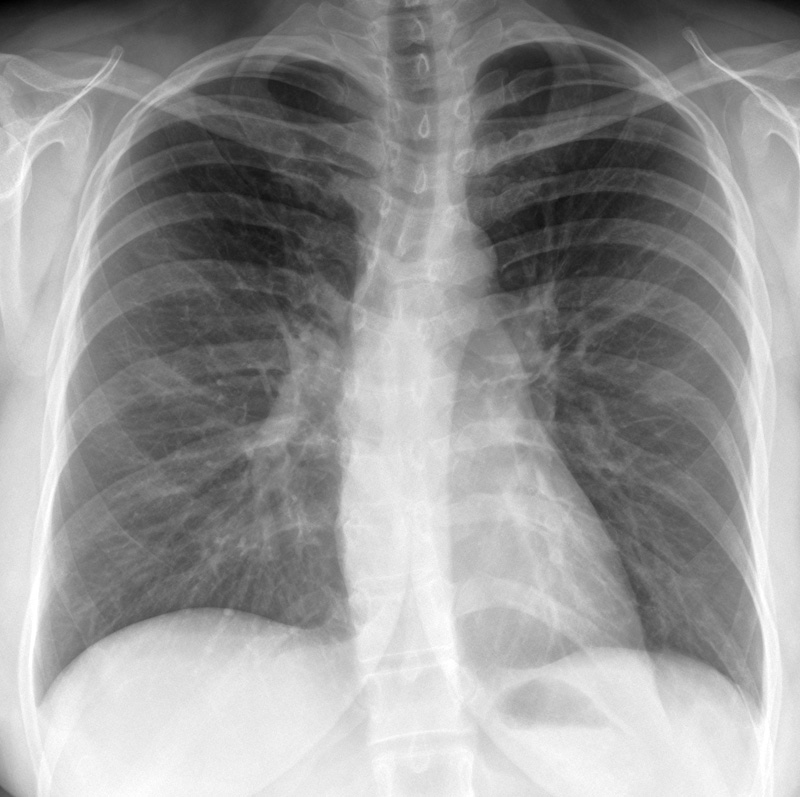
Figure 1. Frontal chest radiography at presentation shows normal heart size, clear lungs, and no pleural abnormality.
Which of the following statements regarding this chest radiograph is accurate? (click on the correct answer to be directed to the first of twelve additional pages)
- Frontal chest radiography shows normal findings
- Frontal chest radiography shows mild cardiomegaly
- Frontal chest radiography shows mediastinal lymphadenopathy
- Frontal chest radiography shows pleural effusion
- Frontal chest radiography shows numerous small nodules