
May 2021 Imaging Case of the Month: A Growing Indeterminate Solitary Nodule
Kenneth K. Sakata, MD‡
Natalya Azadeh, MD, MPH‡
Maxwell Smith, MD↑
Michael B. Gotway, MD†
°Department of Medicine, Mayo Clinic Arizona
↑Department of Laboratory Medicine and Pathology, Mayo Clinic Arizona
‡Division of Pulmonary and Critical Care Medicine, Mayo Clinic Arizona
†Department of Radiology, Mayo Clinic, Arizona
5777 East Mayo Boulevard
Phoenix, Arizona 85054
A 58-year-old woman with a history of orthotopic heart transplant, performed for Adriamycin-induced cardiomyopathy, treated with mycophenolate and tacrolimus, presented for routine interval follow up. The patient’s past medical history was significant for follicular thyroid carcinoma treated with total thyroidectomy and bilateral breast carcinoma in remission as well as hypothyroidism and type II diabetes mellitus. In addition to tacrolimus and mycophenolate, the patient’s medications included aspirin, insulin, itraconazole (for anti-fungal prophylaxis), levothyroxine, prednisone (tapering since transplant), and valganciclovir. The patient recently complained of rhinorrhea and cough productive of brown-tinged sputum, improving over the previous 2 weeks; she denied fever, chills, shortness of breath, night sweats chest pain, or gastrointestinal symptoms.
Physical examination showed the patient to be afebrile with normal heart and respiratory rates and blood pressure. Her room air oxygen saturation was 99%.
The patient’s complete blood count and serum chemistries showed largely normal values, with the white blood cell count at the upper normal at 9.7 x 10⁹ /L (normal, 4-10 x 109 /L). Her liver function testing and renal function testing parameters were also within normal limits. Echocardiography showed normal left ventricular systolic function. The patient underwent frontal chest radiography (Figure 1).
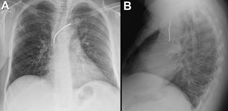
Figure 1. Frontal chest radiography.
Which of the following represents an appropriate interpretation of her frontal chest radiograph? (Click on the correct answer to be directed to the second of nine pages).
- Frontal chest radiography shows a right pleural effusion
- Frontal chest radiograph shows a left apical nodule
- Frontal chest radiography shows multifocal consolidation
- Frontal chest radiography shows peribronchial and mediastinal lymphadenopathy
- Frontal chest radiography shows cardiomegaly
Cite as: Kim JHJ, Sakata KK, Azadeh N, Smith M, Gotway MB. May 2021 Imaging Case of the Month: A Growing Indeterminate Solitary Nodule. Southwest J Pulm Crit Care Med. 2021;229(5):88-99. doi: https://doi.org/10.13175/swjpcc013-21 PDF
 Post a Comment
Reader Comments