Robert W. Viggiano, MD*
Michael B. Gotway, MD**
*Pulmonary Department and **Department of Radiology
Mayo Clinic Arizona
Scottsdale, AZ USA
Clinical History: A 65-year-old non-smoking man with a past medical history significant for hyperlipidemia, hypertension, coronary artery disease, and pacemaker placement, presented for a routine medical evaluation.
The patient was allergic to penicillin, and his list of medications included aspirin, a diuretic, an ACE inhibitor, and a statin, in addition to over-the-counter vitamin supplements. Laboratory evaluation showed a normal complete blood count, electrolyte panel, and liver function tests. Frontal and chest radiography (Figure 1) was performed.
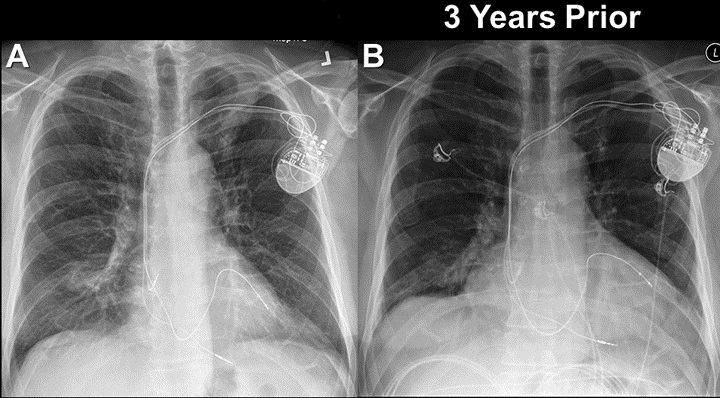
Figure 1. Frontal chest radiography performed at presentation (A) and three years earlier (B).
Which of the following represents the most accurate assessment of the frontal chest imaging findings? (Click on the correct answer to proceed to the second of ten pages)
- Chest frontal imaging shows a mediastinal mass
- Chest frontal imaging shows bilateral peribronchial and mediastinal lymph node enlargement
- Chest frontal imaging shows bilateral pleural fluid collections
- Chest frontal imaging shows focal masses
- Chest frontal imaging shows reduced lung volumes with basilar fibrotic changes
Cite as: Viggiano RW, Gotway MB. April 2018 imaging case of the month. Southwest J Pulm Crit Care. 2018;16(4):194-205. doi: https://doi.org/10.13175/swjpcc056-18 PDF