Courtney M. Tomblinson, MD and Michael B. Gotway, MD
Department of Radiology
Mayo Clinic Arizona
Scottsdale, Arizona USA
Clinical History: A 69-year-old man presented with long-standing complaints of dyspnea, progressing to dyspnea at rest, associated with some dysphagia to solids. He also noted symptoms consistent with exertional stertor (a respiratory sound characterized by heavy snoring or gasping). His past medical history was remarkable only for hypertension controlled with medication.
Laboratory data, include white blood cell count, coagulation profile, and serum chemistries were within normal limits. Oxygen saturation on room air was normal.
Frontal chest radiography (Figure 1) was performed.
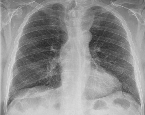
Figure 1: Frontal chest radiography.
Which of the following statements regarding the chest radiograph is most accurate? (Click on the correct answer to proceed to the second of nine pages)
- Frontal chest radiography shows a cavitary lung mass
- Frontal chest radiography shows an abnormal mediastinal contour
- Frontal chest radiography shows multiple small nodules
- Frontal chest radiography shows peribronchial and mediastinal lymphadenopathy
- Frontal chest radiography shows pleural effusion
Cite as: Tomblinson CM, Gotway MB. March 2017 imaging case of the month. Southwest J Pulm Crit Care. 2017;14(3):104-16. doi: https://doi.org/10.13175/swjpcc029-17 PDF