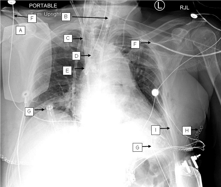
Figure 1. ICU portable chest x-ray. A: cardioversion pads. B: oro-gastric tube. C: right internal jugular dialysis catheter. D: endotracheal tube. E: left internnal jugular central venous catheter, incidentally seen terminating within the azygous vein. F: external EKG lead. G: chest tubes. H: staples along the thoracotomy incision. I: left lower lobe atelectasis and small pleural effusion.
A chest x-ray is probably the most commonly obtained radiographic image in the intensive care unit (ICU). Although not supported by evidence and recommended against, daily chest x-rays, especially in the intubated patients, are done in many ICUs (1,2). Multiple hardware placed for the support of the patient need to be identified for placement, position and potential complications. These can make reading a radiograph challenging specially the mediastinum. The accompanied radiograph serves as an example of an “ICU chest x-ray” with multiple “tube and lines”.
Janet Campion MD and Bhupinder Natt MD
Division of Pulmonary, Allergy, Critical Care and Sleep
Banner-University Medical Center, Tucson (AZ)
References
- Oba Y, Zaza T. Abandoning daily routine chest radiography in the intensive care unit: meta-analysis. Radiology. 2010 May;255(2):386-95. [CrossRef] [PubMed]
- http://www.choosingwisely.org/wp-content/uploads/2015/02/SCCM-Choosing-Wisely-List.pdf
Cite as: Campion J, Natt B. Medical image of the week: ICU chest x-ray. Southwest J Pulm Crit Care. 2017;14(1):39. doi: https://doi.org/10.13175/swjpcc007-17 PDF