
Correct!
Answer 3. Multifocal reticulonodular opacities
The frontal chest radiograph in figure 1 shows mid and lower lung reticular and nodular opacities. No pleural effusion or consolidation is present. These findings are very non-specific and could be caused by numerous processes.
Thoracic CT (Figure 2) was performed.
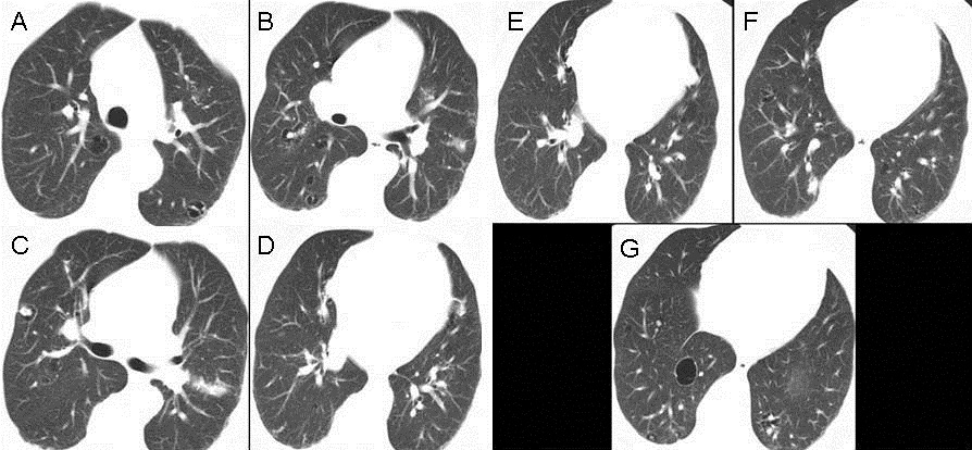
Figure 2. Thoracic CT lung. Lung windows.
Click here for axial thoracic CT scan using lung windows.
Click here for axial thoracic CT scan using soft tissue windows.
Characterize the thoracic CT findings. Which of the following is the LEAST likely diagnostic consideration for the thoracic CT findings.