Correct!
4. 1 and 3
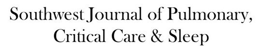
The spirometry on the PFTs show very severe obstruction without reversibility. The reduced forced vital capacity (FVC) suggests the possibility of restriction in addition. The total lung capacity is normal but the residual volume is markedly elevated suggesting the possibility of a combined obstruction and restriction. The diffusion is moderately reduced at 46% of predicted. The thoracic CT shows septal thickening with mosaicism, extensive lower lung bronchiectasis with mucus plugging, tree-in-bud nodularity, signet rings and mediastinal lymphadenopathy (Figure 3).

Figure 3. Thoracic CT showing septal thickening (arrows in A and B), signet rings (circle in C) and enlarged mediastinal lymph nodes (starred in D).
Which of the following are diagnostic considerations? (Click on the correct answer to be directed to the third of six pages)