
Correct!
1. Chest radiography shows a vague solitary pulmonary opacity
The frontal radiograph shows a vague nodular opacity overlying the medial right base, perhaps with some streaky opacity just superior to the nodular opacity (Figure 2).
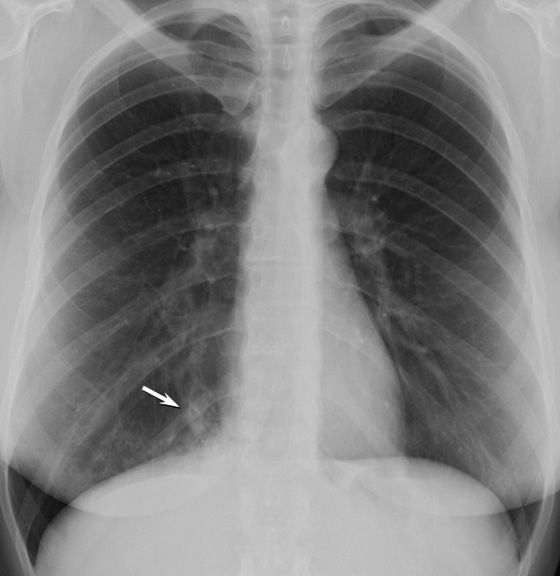
Figure 2. Frontal chest radiography shows a poorly defined nodular opacity (arrow) projected over the medial right lung base. No pleural abnormality or lymph node enlargement is present and the left lung is clear.
The lungs appear relatively clear otherwise. There is no evidence of cavitation, the heart size is normal, the lung volumes are normal, and there is no evidence of fibrotic disease.
At this point, which of the following represents the most appropriate step in this patient’s management? (Click on the correct answer to be directed to the third of sixteen pages)