
Correct!
6. 2 and 4
The thoracic CT shows multiple lung cysts. Rupture of one of these cysts likely led to her pneumothorax. In the setting of cystic lung disease, in order to obtain definitive diagnosis, one should obtain confirmation of the specific type of cystic lung disease, as this will dictate treatment (1,2). Additionally, as she will be at increased risk for recurrent pneumothoraces, consideration should be given to pleurodesis to reduce to chance of recurrence.
The patient underwent VATS with open lung biopsy (Figure 3).
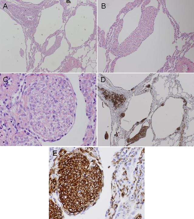
Figure 3. Panel A: 40 x H & E showing foci of smooth muscle cell infiltration of lung parenchyma, airways, lymphatics, and blood vessels associated with thin-walled cystic changes. Panel B: 100 x H & E. Panel C: 400 x H & E showing small spindle shaped cells and cuboid epithelial cells. Panel D: 40 x smooth muscle actin stain (SMA). The cells stain positive to SMA and human melanoma-black 45 stain. Panel E: 400 x SMA stain.
What is the diagnosis? (Click on the correct answer to proceed to the third of five panels)