Correct!
4. An intracavitary mass superimposed on a background of interstitial abnormality
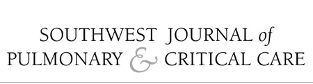
Frontal and lateral chest radiograph shows a mid- and upper lung predominant linear and reticular abnormalities with architectural distortion. A nodule (arrow) is seen projected over the right upper lung, and appears to reside within a thin-walled cavity (arrowheads).
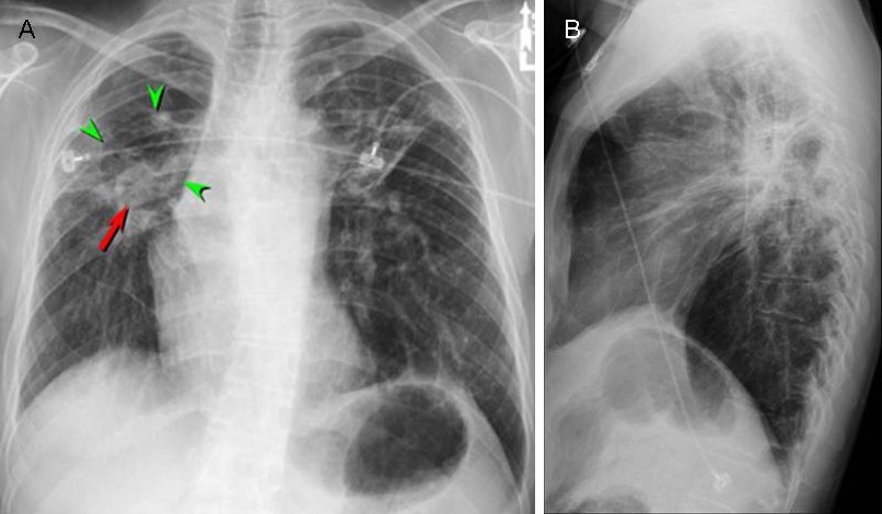
Clinical Course: The patient underwent thoracic CT (Figures 2A and 2B).
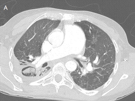
Figures 2A and B: Axial (A) and coronal (B) thoracic CT
Click here for a movie of axial CT images Click here for a movie of coronal CT images
What is the main finding on the thoracic CT?