Search Journal-type in search term and press enter
Southwest Pulmonary and Critical Care Fellowships
In Memoriam

Imaging

Last 50 Imaging Postings

(Click on title to be directed to posting, most recent listed first)

April 2024 Medical Image of the Month: Wind Instruments Player Exhibiting
   Exceptional Pulmonary Function
March 2024 Medical Image of the Month: Sputum Cytology in Patients with
   Suspected Lung Malignancy Presenting with Acute Hypoxic Respiratory
   Failure
February 2024 Medical Image of the Month: Pulmonary Alveolar Proteinosis
   in Myelodysplastic Syndrome
February 2024 Imaging Case of the Month: Connecting Some Unusual Dots
January 2024 Medical Image of the Month: Polyangiitis Overlap Syndrome
   (POS) Mimicking Fungal Pneumonia 
December 2023 Medical Image of the Month: Metastatic Pulmonary
   Calcifications in End-Stage Renal Disease 
November 2023 Medical Image of the Month: Obstructive Uropathy
   Extremis
November 2023 Imaging Case of the Month: A Crazy Association
October 2023 Medical Image of the Month: Swyer-James-MacLeod
   Syndrome
September 2023 Medical Image of the Month: Aspergillus Presenting as a
   Pulmonary Nodule in an Immunocompetent Patient
August 2023 Medical Image of the Month: Cannonball Metastases from
   Metastatic Melanoma
August 2023 Imaging Case of the Month: Chew Your Food Carefully
July 2023 Medical Image of the Month: Primary Tracheal Lymphoma
June 2023 Medical Image of the Month: Solitary Fibrous Tumor of the Pleura
May 2023 Medical Image of the Month: Methamphetamine Inhalation
   Leading to Cavitary Pneumonia and Pleural Complications
April 2023 Medical Image of the Month: Atrial Myxoma in the setting of
   Raynaud’s Phenomenon: Early Echocardiography and Management of
   Thrombotic Disease
April 2023 Imaging Case of the Month: Large Impact from a Small Lesion
March 2023 Medical Image of the Month: Spontaneous Pneumomediastinum
   as a Complication of Marijuana Smoking Due to Müller's Maneuvers
February 2023 Medical Image of the Month: Reversed Halo Sign in the
   Setting of a Neutropenic Patient with Angioinvasive Pulmonary
   Zygomycosis
January 2023 Medical Image of the Month: Abnormal Sleep Study and PFT
   with Supine Challenge Related to Idiopathic Hemidiaphragmatic Paralysis
December 2022 Medical Image of the Month: Bronchoesophageal Fistula in
   the Setting of Pulmonary Actinomycosis
November 2022 Medical Image of the Month: COVID-19 Infection
   Presenting as Spontaneous Subcapsular Hematoma of the Kidney
November 2022 Imaging Case of the Month: Out of Place in the Thorax
October 2022 Medical Image of the Month: Infected Dasatinib Induced
   Chylothorax-The First Reported Case 
September 2022 Medical Image of the Month: Epiglottic Calcification
Medical Image of the Month: An Unexpected Cause of Chronic Cough
August 2022 Imaging Case of the Month: It’s All About Location
July 2022 Medical Image of the Month: Pulmonary Nodule in the
   Setting of Pyoderma Gangrenosum (PG) 
June 2022 Medical Image of the Month: A Hard Image to Swallow
May 2022 Medical Image of the Month: Pectus Excavatum
May 2022 Imaging Case of the Month: Asymmetric Apical Opacity–
   Diagnostic Considerations
April 2022 Medical Image of the Month: COVID Pericarditis
March 2022 Medical Image of the Month: Pulmonary Nodules in the
   Setting of Diffuse Idiopathic Pulmonary NeuroEndocrine Cell Hyperplasia
   (DIPNECH) 
February 2022 Medical Image of the Month: Multifocal Micronodular
   Pneumocyte Hyperplasia in the Setting of Tuberous Sclerosis
February 2022 Imaging Case of the Month: Between A Rock and a
   Hard Place
January 2022 Medical Image of the Month: Bronchial Obstruction
   Due to Pledget in Airway Following Foregut Cyst Resection
December 2021 Medical Image of the Month: Aspirated Dental Implant
   Medical Image of the Month: Cavitating Pseudomonas
   aeruginosa Pneumonia
November 2021 Imaging Case of the Month: Let’s Not Dance
   the Twist
Medical Image of the Month: COVID-19-Associated Pulmonary
   Aspergillosis in a Post-Liver Transplant Patient
Medical Image of the Month: Stercoral Colitis
Medical Image of the Month: Bleomycin-Induced Pulmonary Fibrosis
   in a Patient with Lymphoma
August 2021 Imaging Case of the Month: Unilateral Peripheral Lung
   Opacity
Medical Image of the Month: Hepatic Abscess Secondary to Diverticulitis
   Resulting in Sepsis
Medical Image of the Month: Metastatic Spindle Cell Carcinoma of the
   Breast
Medical Image of the Month: Perforated Gangrenous Cholecystitis
May 2021 Imaging Case of the Month: A Growing Indeterminate Solitary
   Nodule
Medical Image of the Month: Severe Acute Respiratory Distress
   Syndrome and Embolic Strokes from Polymethylmethacrylate
   (PMMA) Embolization
Medical Image of the Month: Pulmonary Aspergillus Overlap Syndrome
   Presenting with ABPA, Multiple Bilateral Aspergillomas
Medical Image of the Month: Diffuse White Matter Microhemorrhages
   Secondary to SARS-CoV-2 (COVID-19) Infection
February 2021 Imaging Case of the Month: An Indeterminate Solitary
   Nodule
Medical Image of the Month: Mucinous Adenocarcinoma of the Lung
   Mimicking Pneumonia
Medical Image of the Month: Superior Vena Cava Syndrome
Medical Image of the Month: Buffalo Chest Identified at the Time of
   Lung Nodule Biopsy
November 2020 Imaging Case of the Month: Cause and Effect?
Medical Image of the Month: Severe Left Ventricular Hypertrophy
Medical Image of the Month: Glioblastoma Multiforme


For complete imaging listings click here

Those who care for patients with pulmonary, critical care or sleep disorders rely heavily on chest radiology and pathology to determine diagnoses. The Southwest Journal of Pulmonary and Critical Care publishes case-based articles with characteristic chest imaging and related pathology. The editor of this section will oversee and coordinate the publication of a core of the most important chest imaging topics. In doing so, they encourage the submission of unsolicited manuscripts. It cannot be overemphasized that both radiologic and pathologic images must be of excellent quality. As a rule, 600 DPI is sufficient for radiographic and pathologic images. Taking pictures of plain chest radiographs and CT scans with a digital camera is strongly discouraged. The figures should be cited in the text and numbered consecutively. The stain used for pathology specimens and magnification should be mentioned in the figure legend. Those who care for patients with pulmonary, critical care or sleep disorders rely heavily on chest radiology and pathology to determine diagnoses. The Southwest Journal of Pulmonary and Critical Care publishes case-based articles with characteristic chest imaging and related pathology. The editor of this section will oversee and coordinate the publication of a core of the most important chest imaging topics. In doing so, they encourage the submission of unsolicited manuscripts. It cannot be overemphasized that both radiologic and pathologic images must be of excellent quality. As a rule, 600 DPI is sufficient for radiographic and pathologic images. Taking pictures of plain chest radiographs and CT scans with a digital camera is strongly discouraged. The figures should be cited in the text and numbered consecutively. The stain used for pathology specimens and magnification should be mentioned in the figure legend.

-------------------------------------------------------------------------------------------  

Wednesday
Nov132013

Medical Image of the Week: Migration of Fractured PICC Line

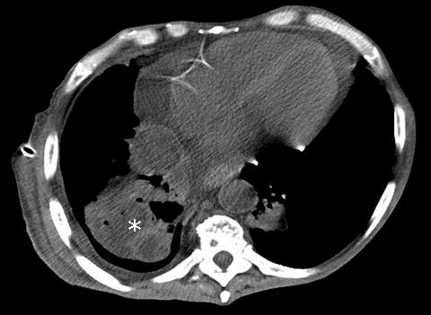
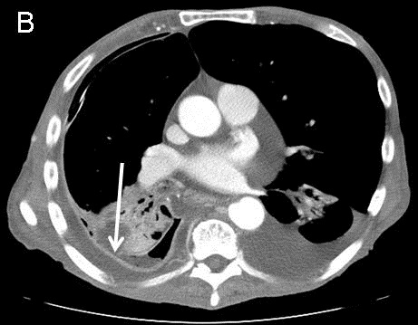
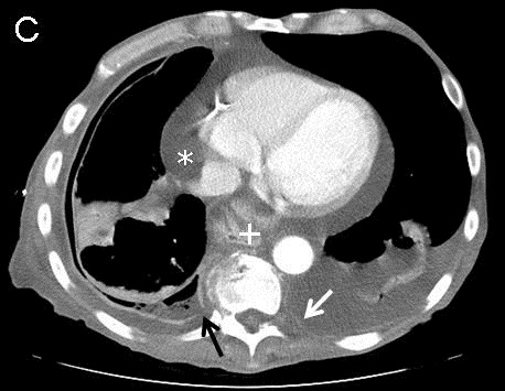
Figure 1. Chest radiograph showing catheter fragment coiled within the main pulmonary artery with the tips extending to the peripheral pulmonary vasculature (arrow).

A 29 year old man had a peripherally inserted central catheter (PICC, 60 cm, 4 Fr, single lumen Groshong), placed at a rural hospital via right brachial vein approach for long term antibiotic therapy for an infected diabetic foot. Two days later, nursing staff noticed difficulty in flushing the PICC line. A chest X-ray was obtained which showed fracture of a portion of the PICC line with migration to the pulmonary vasculature (Figure 1). The broken catheter was successfully removed in a two-step process using a 6 x10 mm EN Snare® from the left pulmonary artery. The patient, however remained asymptomatic and without any arrhythmias on the telemonitor. Intravascular migration and subsequent malposition of PICC lines are usually due to their pinch off, small size, as well as changes in intrathoracic pressure and body position (1).

Santhosh G. John MD, Paola Devis MD

University of Arizona

Tucson, AZ

Reference

  1. Zhang Y, Jiang H, Wei L, Yu H. Persistent hiccup Caused by peripherally inserted central catheter migration. J Anesth. 2011;25(4):625-6. [CrossRef] [PubMed]

Reference as: John SG, Devis P. Medical image of the week: migration of fractured PICC line. Southwest J Pulm Crit Care. 2013;7(5): . doi: http://dx.doi.org/10.13175/swjpcc151-13 PDF

Wednesday
Nov062013

Medical Image of the Week: Empyema

Figure 1. Admission thoracic CT showing a low density collection in the right lung likely from necrosis (*).


Figure 2. Selected views from the thoracic CT obtained 1 week after admission. Panel A: Architectural distortion (white circle) suggests necrotizing pneumonia. Note fluid within the bronchus intermedius (*). Panel B: Defect in visceral pleura (arrow) with decompression of parenchymal necrosis into the pleural space.  Note enhancement and thickening of both pleural layers. Panel C: Defect in parietal pleura (black arrow) with fluid extending into the extrapleural space. Pericardial effusion (*), new left pleural effusion and left parietal pleura enhancement (white arrow) suggests spread of infection. Incidental hiatal hernia (+).


A 71 year-old man with chronic obstructive pulmonary disease (COPD) presents to the emergency department complaining of dyspnea after recent admission for pneumonia. Chest CT shows a low density collection in the right lung suggesting necrosis (Figure 1). A CT obtained 1 week after admission (Figure 2) shows progression to empyema.

Management of empyema can be difficult. If the fluid cannot be removed with a therapeutic thoracentesis, a chest tube should be inserted and consideration be given to the intrapleural instillation of fibrinolytics (1). If the loculated effusion persists, the patient should be subjected to video-assisted thoracoscopic surgery. If the lung cannot be expanded with this procedure, a full thoracotomy with decortication should be performed. The definitive procedure should be performed within fourteen days.

Jason R. Young MD and David L. August, MD

Department of Radiology

Maricopa Integrated Health System

Phoenix, AZ

Reference

  1. Light RW. Parapneumonic effusions and empyema. Proc Am Thorac Soc. 2006;3(1):75-80. [CrossRef] [PubMed]

Reference as: Young JR, August DL. Medical image of the week: empyema. Southwest J Pulm Crit Care. 2013;7(5):300-1. http://dx.doi.org/10.13175/swjpcc143-13 PDF