Michael B. Gotway, MD
Department of Radiology
Mayo Clinic Arizona
Scottsdale, AZ USA
Clinical History: A 51-year-old previously healthy man presented with complaints of increasing headache frequency and severity. The patient noted headaches in the past, but that the frequency of these headaches, which he referred to as “migraines,” had been increasing in recent months. The patient does note some auras with the headaches.
The patient reported a history of pneumonia in the past, but denied recurrent pneumonias. The only medication the patient takes was ibuprofen, for his headaches; he denied allergies. The patient’s past surgical history was remarkable only for a right inguinal hernia repair, a right Achilles tendon injury repair, and surgical removal of a palpable left thigh mass, ultimately shown to represent scar tissue. The patient smoked 1-8 cigarettes / day for 35 years, quitting one year earlier.
The patient’s physical examination was remarkable for obesity (BMI= 30.4). His vital signs were within the normal range. A few reddish rounded spots were noted on his lower lip, but no other abnormalities were noted at physical examination.
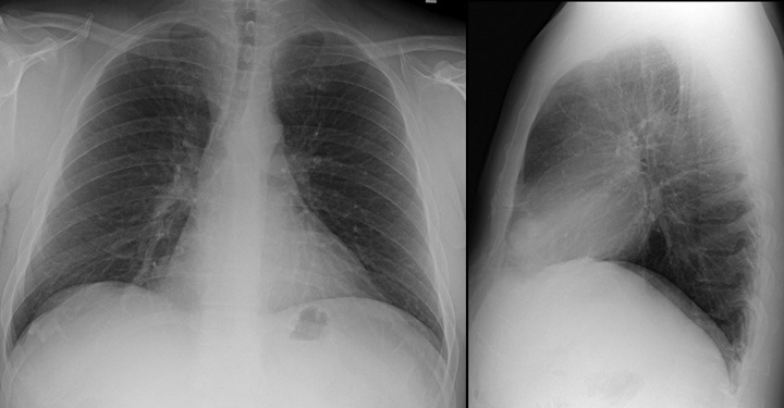
Basic laboratory data, including a complete blood count, electrolyte panel, B12 and folate levels, a C-reactive protein level, and liver function studies were all within the normal range. Mild hypercholesterolemia was noted. An electrocardiogram revealed normal findings. As part of a routine office visit, frontal and lateral chest radiography (Figure 1) was performed.
Figure 1. Frontal and lateral chest radiography
Which of the following statements regarding the chest radiograph is most accurate? (Click on the correct answer to proceed to the second of ten pages)
Cite as: Gotway MB. August 2019 imaging case of the month: a 52-year-old man with a headache. Southwest J Pulm Crit Care. 2019;19(2):52-64. doi: https://doi.org/10.13175/swjpcc052-19 PDF