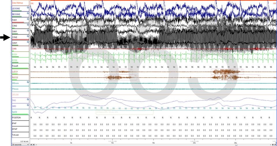
Figure 1. Thirty second polysomnogram epoch showing artifact in lead O1M2 (black arrow).
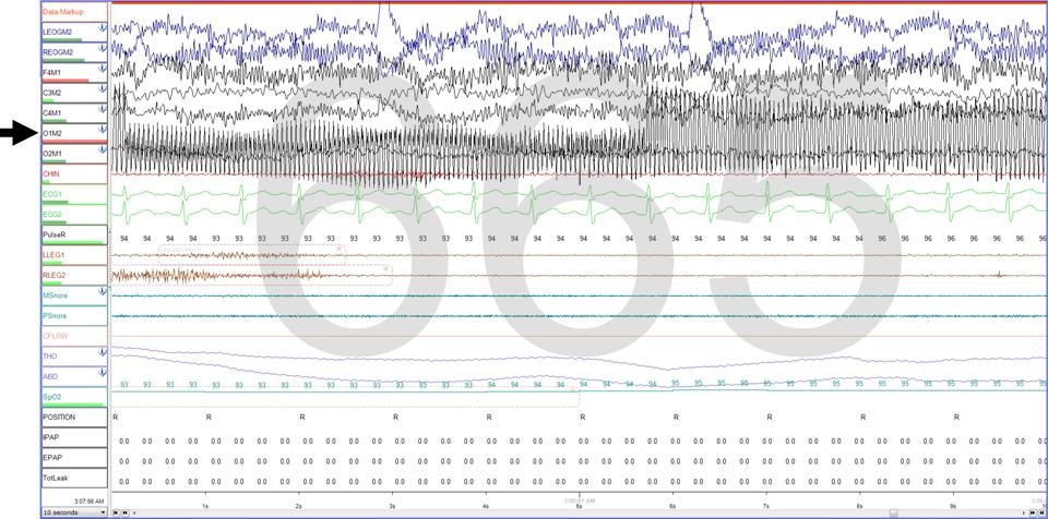
Figure 1. Ten second polysomnogram epoch showing artifact in lead O1M2 (black arrow).
A 54 year-old man with a past medical history of attention deficit hyperactivity disorder (ADHD), low back pain, and paroxysmal supraventricular tachycardia presented to the sleep laboratory for evaluation of sleep disordered breathing. Pertinent medications include fluoxetine, ambien, and clonazepam. His Epworth sleepiness score was 18. He had a total sleep time of 12 min. On the night of his sleep study, the patient was restless and repeatedly changed positions in bed.
Figures 1 and 2 show the artifact determined to be lead displacement of O1M2 after the patient shifted in bed, inadvertently removing one of his scalp electrodes. The sine waves are 60 Hz in frequency. Once the problem was identified, the lead was quickly replaced to its proper position.
Jared Bartell1, Safal Shetty, MD1,2, and John D. Roehrs, MD1,2
1University of Arizona Medical Center
2Southern Arizona VA Health Care System
Tucson, AZ
Reference as: Bartell J, Shetty S, Roehrs JD. Medical image of the week: polysomnogram artifact. Southwest J Pulm Crit Care. 2015;10(2):95-6. doi: http://dx.doi.org/10.13175/swjpcc014-15 PDF