Michael B. Gotway, MD
Department of Radiology
Mayo Clinic Arizona
Scottsdale, AZ
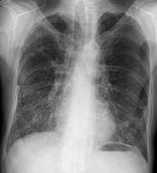
Clinical History: A 60-year-old man presented with a history of weight loss and dysphagia for about 2 weeks duration. There was a possible history of asthma accompanied by ongoing shortness of breath first noticed nearly 2 years ago. Frontal chest radiography (Figure 1) was performed.

Figure 1. Frontal chest radiography.
Which of the following statements regarding the chest radiograph is most accurate? (Choose the correct answer to move to the next panel)
- The chest radiograph shows a mass
- The chest radiograph shows hilar and mediastinal lymph node enlargement
- The chest radiograph shows multifocal consolidation
- The chest radiograph shows multifocal, somewhat basal predominant linear opacities suggesting fibrosis
- The chest radiograph shows multiple nodules
Reference as: Gotway MB. February 2014 imaging case of the month. Southwest J Pulm Crit Care. 2014;8(2):88-95. doi: http://dx.doi.org/10.13175/swjpcc010-14 PDF