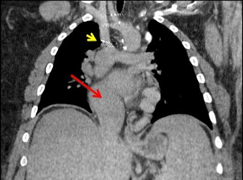
Figure 1. Thoracic CT scan showing Fontan anatomy, with the superior vena cava (SVC) connected to the pulmonary arteries (yellow arrow) and a single atrium and ventricle (red arrow).

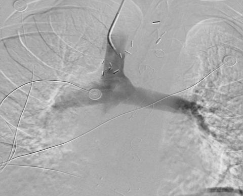
Figure 2. SVC venography shows SVC connected to the pulmonary artery.
A 25-year-old man with a history of transposition of the great vessels (L-TGA) was admitted for persistent hemoptysis. He had a history of a double inlet left ventricle, pulmonary hypertension and was postoperative for a Fontan procedure completed at age of 2. No anatomical source for the hemoptysis was found. A thoracic CT showed the Fontan anatomy: SVC connected to the pulmonary artery as per the Glenn connection (IVC drained to right pulmonary artery through the Fontan pathway) and a single ventricle and atrium (Figure 1). SVC venography showed the SVC connected to the pulmonary artery (Figure 2). The hemoptysis resolved after started sidenafil and bosentan for pulmonary hypertension.
Mohammed Alzoubaidi MD, Carmen Luraschi Monjagatta MD, Maria Tumanik DO, Naomi Jean Young MD
University of Arizona
Department of Pulmonary and Critical Care Medicine
Internal Medicine, South Campus.
Family Medicine, South Campus
Tucson, AZ
Reference as: Alzoubaidi M, Monjagatta CL, Tumanik M, Young NJ. Medical image of the week: Fontan procedure. Southwest J Pulm Crit Care. 2013;7(2):112-3. doi: http://dx.doi.org/10.13175/swjpcc114-13 PDF