Michael B. Gotway, MD
Department of Radiology
Mayo Clinic Arizona
Scottsdale, AZ
Clinical History
A 64-year-old woman with a history of multiple sclerosis (wheelchair-bound), neurogenic bladder, and a number of other chronic medical conditions, presented with complaints of non-radiating neck pain without tingling or numbness. The patient also reported mild subjective fever and occasional nausea, but denied shortness of breath. Frontal and lateral chest radiography (Figure 1) was performed.
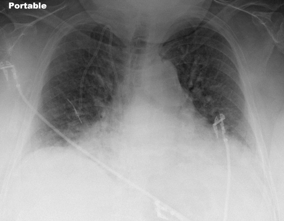
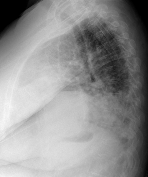 Figure 1. Frontal (Panel A) and lateral (Panel B) chest x-ray.
Which of the following statements regarding the chest radiograph is most accurate?
- The chest radiograph shows bibasilar consolidation
- The chest radiograph shows large lung volumes with cystic change
- The chest radiograph shows multiple nodules
- The chest radiograph shows no abnormalities
- The chest radiograph shows symmetrical bilateral pleural effusions
Reference as: Gotway MB. July 2013 imaging case of the month. Southwest J Pulm Crit Care. 20130.;7(1):17-24. doi: http://dx.doi.org/10.13175/swjpcc087-13 PDF