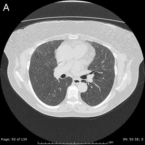
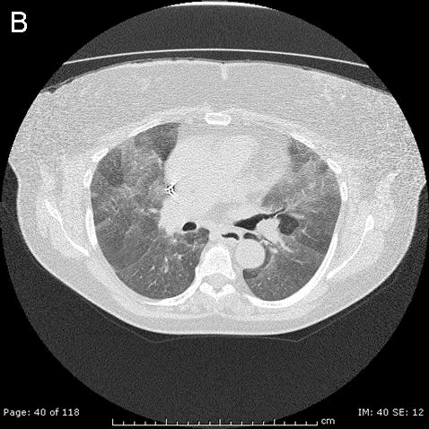
A 66 year old female presented with cough, fever and marked shortness of breath. Infectious work up was found to be negative. An inspiratory high resolution thoracic CT (HRCT) image (A) shows faint groundglass and mosaic lung attenuation with subtle centrilobular ill-defined nodules. However, an image obtained on expiration (B) shows more obvious mosaic attenuation which suggesting air-trapping. Due to progressive dyspnea, a lung biopsy was performed and revealed a bronchiolocentric cellular interstitial pneumonia with non-caseating granuloma consistent with subacute hypersensitivity pneumonitis.
Veronica A. Arteaga, MD and Kenneth S. Knox, MD
Divisions of Thoracic Imaging and Pulmonary/Critical Care Medicine
University of Arizona
Tucson, Arizona
Reference as: Arteaga VA, Knox KS. Medical image of the week: expiratory imaging accentuates mosaic attenuation. Southwest J Pulm Crit Care. 2013;6(5):245. PDF