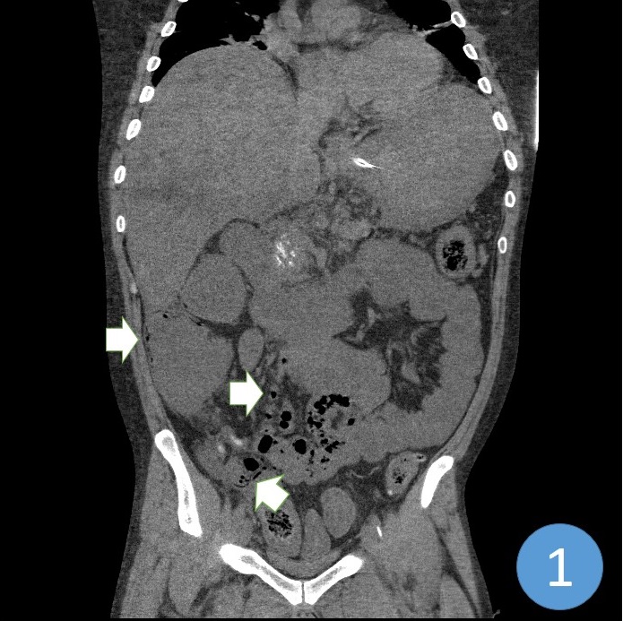
Figure 1. Coronal view of a non-contrast CT scan demonstrating pneumatosis intestinalis of the distal small bowel, ascending colon, and superior mesenteric vein (arrows).
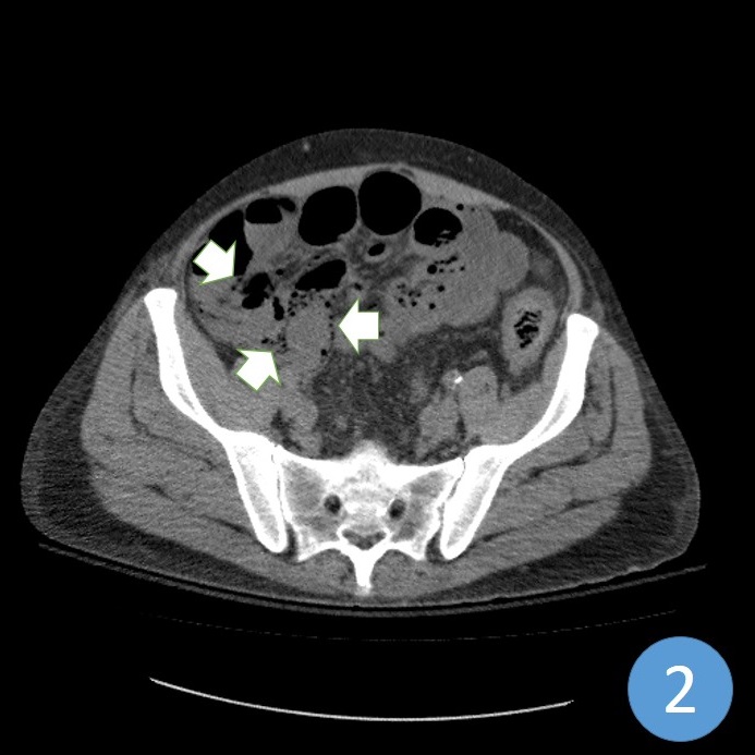
Figure 2. Transverse cross-section of severe pneumatosis secondary to acute blood loss intestinal ischemia (arrows).
The patient was a 32 year-old male with a past medical history significant for end stage liver disease secondary to severe alcoholism who was found with an altered mental status. In the emergency department, the patient divulged he had been throwing up blood clots in the preceding days. Shortly into his presentation he began throwing up voluminous bright red blood. Initial hemoglobin concentration was 2.8 mg/dL. CT scan of the abdomen revealed pneumatosis within the ascending colon, small bowel, and mesenteric veins. Despite massive transfusion efforts and two episodes of successful cardiac resuscitation the patient expired.
Seth Assar, MD; Herman Solorzano; Ishna Poojari, MD; Maria del Carmen Luraschi Monjagatta, MD
The University of Arizona College of Medicine at South Campus, Tucson, Arizona
References
- Pieterse AS, Leong AS, Rowland R. The mucosal changes and pathogenesis of pneumatosis cystoides intestinalis. Hum Pathol. 1983;16(7):683-8. [CrossRef]
- Heng Y, Schuffler MD, Haggitt RC, Rohrmann CA. Pneumatosis intestinalis: a review. Am J Gastroenterol. 1995;90(10):1747.[PubMed]
Reference as: Assar S, Solorzano H, Poojari I, Monjagatta MCL. Medical image of the week: pneumatosis intestinalis secondary to massive acute blood loss. Southwest J Pulm Crit Care. 2013;7(4): . doi: http://dx.doi.org/10.13175/swjpcc135-13 PDF