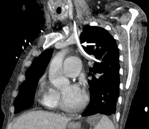
Figure 1. Thoracic CT scan showing the tip of the central line deviating beyond the expected margin of the aortic wall (arrow).
A 51 year old woman was admitted for treatment of acute kidney injury. A dialysis catheter and central line were inserted with ultrasound guidance. Thoracic CT scan (Figure 1) showed the tip of the left central line deviated laterally beyond the expected margin of the aortic wall (arrow). Blood gases were measured showing a partial pressure of oxygen of 154 mm Hg. Arterial line transducer was connected and tracing was consistent with venous system pressures. A CT angiogram revealed the left internal jugular catheter was within the left superior pulmonary vein which was anomalously draining to the left brachiocephalic vein. This explained the arterial-like oxygenation and venous pressure tracing on arterial transducer. The central line was removed without the need of surgical intervention.
Hiram Rivas-Perez MD and Maria Lucarelli MD
Division of Pulmonary, Allergy, Critical Care and Sleep Medicine
Ohio State University
Columbus, Ohio
Reference as: Rivas-Perez H, Lucarelli M. Medical image of the week: anomalous pulmonary venous circulation. Southwest J Pulm Crit Care 2013;6(1):36. PDF