Search Journal-type in search term and press enter
Southwest Pulmonary and Critical Care Fellowships
In Memoriam

Critical Care

Last 50 Critical Care Postings

April 2024 Critical Care Case of the Month: A 53-year-old Man Presenting
   with Fatal Acute Intracranial Hemorrhage and Cryptogenic Disseminated
   Intravascular Coagulopathy 
Delineating Gastrointestinal Dysfunction Variants in Severe Burn Injury
   Cases: A Retrospective Case Series with Literature Review
Doggonit! A Classic Case of Severe Capnocytophaga canimorsus Sepsis
January 2024 Critical Care Case of the Month: I See Tacoma
October 2023 Critical Care Case of the Month: Multi-Drug Resistant
   K. pneumoniae
May 2023 Critical Care Case of the Month: Not a Humerus Case
Essentials of Airway Management: The Best Tools and Positioning for 
   First-Attempt Intubation Success (Review)
March 2023 Critical Care Case of the Month: A Bad Egg
The Effect of Low Dose Dexamethasone on the Reduction of Hypoxaemia
   and Fat Embolism Syndrome After Long Bone Fractures
Unintended Consequence of Jesse’s Law in Arizona Critical Care Medicine
Impact of Cytomegalovirus DNAemia Below the Lower Limit of
Quantification: Impact of Multistate Model in Lung Transplant Recipients
October 2022 Critical Care Case of the Month: A Middle-Aged Couple “Not
   Acting Right”
Point-of-Care Ultrasound and Right Ventricular Strain: Utility in the
   Diagnosis of Pulmonary Embolism
Point of Care Ultrasound Utility in the Setting of Chest Pain: A Case of
   Takotsubo Cardiomyopathy
A Case of Brugada Phenocopy in Adrenal Insufficiency-Related Pericarditis
Effect Of Exogenous Melatonin on the Incidence of Delirium and Its 
   Association with Severity of Illness in Postoperative Surgical ICU Patients
Pediculosis As a Possible Contributor to Community-Acquired MRSA
Bacteremia and Native Mitral Valve Endocarditis
April 2022 Critical Care Case of the Month: Bullous Skin Lesions in
   the ICU
Leadership in Action: A Student-Run Designated Emphasis in
   Healthcare Leadership
MSSA Pericarditis in a Patient with Systemic Lupus
   Erythematosus Flare
January 2022 Critical Care Case of the Month: Ataque Isquémico
   Transitorio in Spanish 
Rapidly Fatal COVID-19-associated Acute Necrotizing
   Encephalopathy in a Previously Healthy 26-year-old Man 
Utility of Endobronchial Valves in a Patient with Bronchopleural Fistula in
   the Setting of COVID-19 Infection: A Case Report and Brief Review
October 2021 Critical Care Case of the Month: Unexpected Post-
   Operative Shock 
Impact of In Situ Education on Management of Cardiac Arrest after
   Cardiac Surgery
A Case and Brief Review of Bilious Ascites and Abdominal Compartment
   Syndrome from Pancreatitis-Induced Post-Roux-En-Y Gastric Remnant
   Leak
Methylene Blue Treatment of Pediatric Patients in the Cardiovascular
   Intensive Care Unit
July 2021 Critical Care Case of the Month: When a Chronic Disease
   Becomes Acute
Arizona Hospitals and Health Systems’ Statewide Collaboration Producing a
   Triage Protocol During the COVID-19 Pandemic
Ultrasound for Critical Care Physicians: Sometimes It’s Better to Be Lucky
   than Smart
High Volume Plasma Exchange in Acute Liver Failure: A Brief Review
April 2021 Critical Care Case of the Month: Abnormal Acid-Base Balance
   in a Post-Partum Woman
First-Attempt Endotracheal Intubation Success Rate Using A Telescoping
   Steel Bougie 
January 2021 Critical Care Case of the Month: A 35-Year-Old Man Found
   Down on the Street
A Case of Athabaskan Brainstem Dysgenesis Syndrome and RSV
   Respiratory Failure
October 2020 Critical Care Case of the Month: Unexplained
   Encephalopathy Following Elective Plastic Surgery
Acute Type A Aortic Dissection in a Young Weightlifter: A Case Study with
   an In-Depth Literature Review
July 2020 Critical Care Case of the Month: Not the Pearl You Were
   Looking For...
Choosing Among Unproven Therapies for the Treatment of Life-Threatening
   COVID-19 Infection: A Clinician’s Opinion from the Bedside
April 2020 Critical Care Case of the Month: Another Emerging Cause
   for Infiltrative Lung Abnormalities
Further COVID-19 Infection Control and Management Recommendations for
   the ICU
COVID-19 Prevention and Control Recommendations for the ICU
   Loperamide Abuse: A Case Report and Brief Review
Single-Use Telescopic Bougie: Case Series
Safety and Efficacy of Lung Recruitment Maneuvers in Pediatric Post-
   Operative Cardiac Patients
January 2020 Critical Care Case of the Month: A Code Post Lung 
   Needle Biopsy
October 2019 Critical Care Case of the Month: Running Naked in the
   Park
Severe Accidental Hypothermia in Phoenix? Active Rewarming Using 
   Thoracic Lavage
Left Ventricular Assist Devices: A Brief Overview


For complete critical care listings click here.

The Southwest Journal of Pulmonary and Critical Care publishes articles directed to those who treat patients in the ICU, CCU and SICU including chest physicians, surgeons, pediatricians, pharmacists/pharmacologists, anesthesiologists, critical care nurses, and other healthcare professionals. Manuscripts may be either basic or clinical original investigations or review articles. Potential authors of review articles are encouraged to contact the editors before submission, however, unsolicited review articles will be considered.

------------------------------------------------------------------------------------

Monday
May012023

May 2023 Critical Care Case of the Month: Not a Humerus Case

Carli S. Ogle1 DO

Billie Bixby2 MD

Janet Campion2 MD

Departments of Family and Community Medicine1 and Internal Medicine2

Banner University Medical Center-South Campus

Tucson, AZ USA


History of Present Illness:

A 57-year-old woman with history of bone disease presented with a 3-day history of cough with thick yellow phlegm and progressive shortness of breath. No fever, chest pain or abdominal pain was noted. In the emergency department, she had SpO2 of 55% on room air, and then 90% on 15L NRB.

Past Medical History/Social History/Family History

  • Bone disease since birth
  • Asthma
  • Severe scoliosis
  • Gastrointestinal reflux disease
  • Cholecystectomy
  • Spinal growth rods
  • Lives in adult care home, supportive family
  • No smoking or alcohol use
  • No illicit drug use
  • There is no family history of any bone disease

Home Medications:

  • Albuterol MDI PRN
  • Alendronate 10mg daily
  • Budesonide nebulizer BID
  • Calcium carbonate BID
  • MVI daily
  • Lisinopril 10mg daily
  • Loratadine 10mg daily
  • Metformin 500mg BID
  • Metoprolol 12.5mg BID
  • Montelukast 10mg daily
  • Naprosyn PRN
  • Omeprazole 20mg daily
  • Simvastatin 10mg daily
  • Tizanidine PRN
  • Vitamin D 2000 IU daily

Allergies:

  • Cefazolin, PCN, Sulfa - all cause anaphylaxis

Physical Examination :

  • Vital signs: BP 135/95, HR 108, RR 36, Temp 37.0 C Noted to desaturate to SpO2 in 70-80s off of Bipap even when on Vapotherm HFNC
  • General: Alert, slightly anxious woman, tachypneic, able to answer questions
  • Skin: No rashes, warm and dry
  • HEENT: No scleral icterus, dry oral mucosa, normal conjunctiva
  • Neck: No elevated JVP or LAD, short length
  • Pulmonary: Diminished breath sounds at bases, no wheezes or crackles
  • Cardiovascular: Tachycardic, regular rhythm without murmur
  • Abdomen: Soft nontender, nondistended, active bowel sounds
  • Extremities: Congenital short upper and lower limb deformities
  • Neurologic: Oriented, fully able to make health care decisions with family at bedside

Laboratory Evaluation:

  • Na 142, K 4.3, CL 100, CO2 29, BUN 15, Cr 0.38, Glu 222
  • WBC 21.9, Hgb 13.6, Hct 42.9, Plt 313 with 83% N, 8% L, 1% E
  • Normal LFTs
  • Lactic acid 2.2
  • Venous Blood Gases (peripheral) on Bipap 10/5, FiO2 90%: pH 7.36, pCO2 58, pO2 55
  • COVID-19 positive

Radiologic Evaluation:

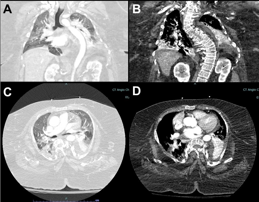
A thoracic CT scan was performed (Figure 1).

Figure 1. Representative images from thoracic CT scan in lung windows (A,C) and soft tissue windows (B,D).

The CT images show all the following except: (Click on the correct answer to be directed to the second of seven pages)

  1. Severe scoliosis
  2. Diffuse ground glass opacities
  3. Right lower lobe consolidation
  4. Pneumothorax
  5. Atelectasis in bilateral lower lobes
Cite as: Ogle CS, Bixby B, Campion J. May 2023 Critical Care Case of the Month: Not a Humerus Case. Southwest J Pulm Crit Care Sleep. 2023;26(5):76-79. doi: https://doi.org/10.13175/swjpccs018-23 PDF


Wednesday
Apr192023

Essentials of Airway Management: The Best Tools and Positioning for First-Attempt Intubation Success

Evan D. Schmitz MD

Pulmonary and Critical Care Medicine

Abstract

Head position during endotracheal intubation affects first-attempt success, as does the different tools available and the location. It is important to be skilled in the operation of a variety of laryngoscopes (video or direct) as well as introducers (plastic/steel stylets and bougies). Difficult airways should always be anticipated and proper preparation such as upper airway assessment performed. The following is a review of endotracheal intubations performed outside of the operating room.

Objectives

  • Discuss how different locations in the hospital can affect endotracheal intubation success.
  • Learn the difference between simple head positioning and the sniffing position and why one should be chosen over the other. MRI images of the head and neck in each position will be reviewed.
  • Learn about different types of laryngoscope blades.
  • Understand the dangers of video laryngoscopy as well as the benefits and when to choose direct laryngoscopy.
  • Define endotracheal intubation first-attempt success.
  • The benefits of using a bougie as opposed to a stylet to increase first-attempt success rate with a review of the supportive literature.
  • Case presentations.

Abbreviations

  • AF – atrial fibrillation
  • ARDS – acute respiratory distress syndrome
  • BiPAP – bilevel positive airway pressure
  • CAD – coronary artery disease
  • COPD – chronic obstructive pulmonary disease
  • Ó – delta
  • DM – diabetes mellitus
  • DVT – deep vein thrombosis
  • ED – emergency department
  • ETT – endotracheal tube
  • FiO2 – fraction of inspired oxygen
  • HFNC – high flow nasal canula
  • HTN – hypertension
  • ICU – intensive care unit
  • LA – laryngeal axis
  • LV – line of vision
  • MRI – magnetic resonance imaging
  • NIDDM – non-insulin dependent diabetes mellitus
  • NRB – non-rebreather mask
  • OA – oral axis
  • OR – operating room
  • OSA – obstructive sleep apnea
  • PA – pharyngeal axis
  • PCO2 – partial pressure of carbon dioxide
  • PE – pulmonary embolism
  • RCA – right coronary artery
  • SpO2 – pulse oximeter oxygen saturation
  • Sz – seizure

Introduction

Ideal positioning can make the difference between a successful endotracheal intubation or death. Many times, intubations are performed in emergency situations, and positioning is not always ideal depending on the type of surface. In the OR, ideal conditions exist regarding adequate supplies and time (1). Conditions can be very different outside of the operating room (OR) especially during a code blue. The average time of intubation is 37 seconds in the emergency department (ED) (2). During the COVID-19 pandemic, intubations were being performed as quickly as 15 seconds in the intensive care unit (ICU) to prevent cardiac arrest in patients with severe adult respiratory distress syndrome (ARDS) (3).

Hospital beds are cumbersome and can cause poor positioning making intubation difficult. If possible, it is always a good idea to have a few towels available to help with head positioning. Towels can be rolled up and placed between the shoulder blades to aid in simple head extension. Towels can also be used to flex the neck on the chest and extend the head on the neck into the sniffing position. Pillows can be added if needed in morbidly obese patients.

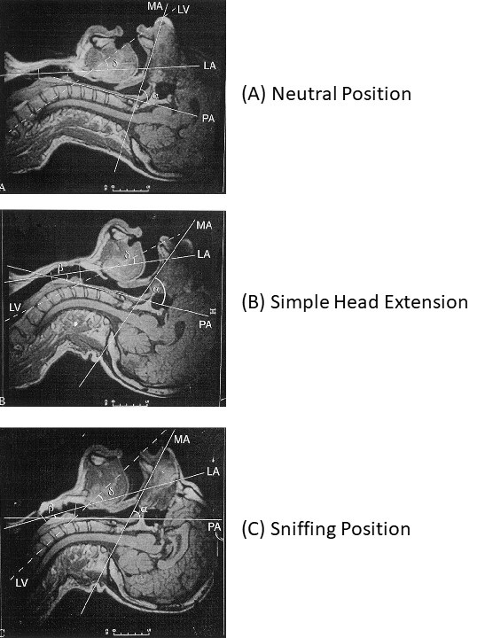
Previous studies published in the Journal of Anesthesia comparing head positioning with regards to line of vision (LV), oral axis (OA), pharyngeal axis (PA), and laryngeal axis (LA) proved that all axes can never be perfectly aligned (Figure 1) (4). The same authors concluded that routine use of the sniffing position appears to provide no significant advantage over simple head extension for tracheal intubation (5).

The sniffing position improved glottic exposure in 18% of patients and worsened it in 11% in comparison with simple head extension in patients intubated in the operating room. Multivariant analysis showed that patients with reduced neck mobility and obesity did better in the sniffing position.

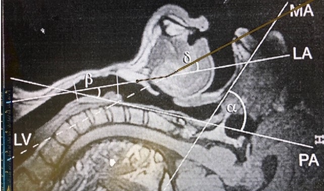
The angle between the LV to the LA, ó, decreases significantly when placed in simple head extension (B) and the sniffing position (C) compared with neutral positioning (A) (Figure 1). In simple head extension ó is the smallest approximating 20o. The smaller the ó, the easier it is to access the glottis. Bougie introducers like the AIROD® telescopic steel bougie with a 20o bend at the proximal end as well as elastic bougies with a coude (bent) tip allow easy transition from the LV to the laryngeal axis LA in simple head extension Figure 2 (6-10).

Figure 1.  Evaluation of the four axes (mouth axis [MA], pharyngeal axis [PA], laryngeal axis [LA], line of vision [LV] and the α, β, and ό angles in the three positions (4).

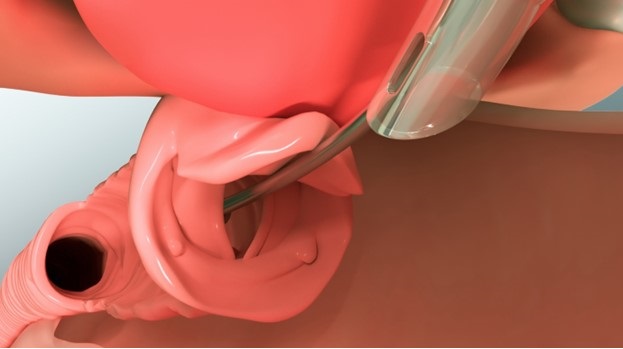
Figure 2. AIROD® aligned perfectly with the laryngeal view (LV) with the head in simple extension. Transition to the laryngeal axis (LA) is easy due to the specialized 20o tip.

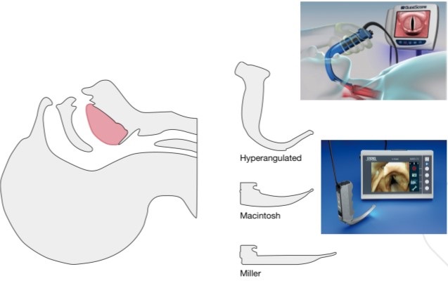
The different video laryngoscopes all offer indirect views of the glottis (Figure 3).


Figure 3. Different types of video and direct laryngoscopes.

For those on C-spine precautions, a hyperangulated Glidescope® or C-MAC® can help with the acute angles involved without the need for significant neck movement. Although video laryngoscopes may improve the view of the glottis because they do not guarantee a direct pathway to the vocal cords, disaster may occur during intubation. Additional tools and expertise should be available immediately because once sedatives and paralytics are given you may no longer be able to ventilate the patient.

In 2017 Baptiste et al. (11) published a study showing that severe life-threatening complications were higher in those ICU patients who were intubated using video laryngoscopy 9.5% vs 2.8% in those who were intubated with direct laryngoscopy with the numbers needed to harm of 14.6. Blood, emesis, secretions, damaged screen, and sudden battery failure can all obscure the video images, complicating intubation with video devices. It is therefore recommended that operators be comfortable using direct laryngoscopes as well as bougies in case of video device failures.

Prior to intubation, airway assessment should be performed to determine whether a difficult airway may be present. If any of the following characteristics are present, then a difficult airway should be expected and precautions taken:

  • Mouth opening < 3.5 cm
  • Thyromental distance < 6.5 cm
  • BMI > 30 kg/m2
  • Amplitude of head and neck movement < 80o
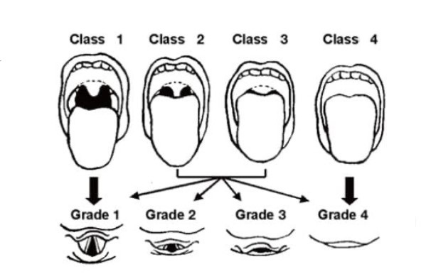
  • Mallampati score > 3
  • Cormack and Lehane classification > 2

Figure 4. Mallampati scores classes 1-4 and Cormack and Lehane classification grades 1-4.

In addition to these measurements, a difficult airway is present if the airway is obstructed by emesis, blood, foreign object or swelling; if the patient has a short neck, large tongue, facial trauma; or if cervical spine immobilization is needed.

Increased complications arise during intubation when a difficult airway is present, especially in an unstable patient. Adverse events related to endotracheal intubation in the ED have been reported at 12% (11). Only 70% of patients intubated in the ICU are successfully intubated upon first-attempt (12). A successful first-attempt intubation is defined as the placement of an endotracheal tube into the trachea upon the initial insertion of the laryngoscope into the oropharynx. If the laryngoscope must be removed and a second-attempt performed, it is considered a failure. Failure to intubate with the first-attempt contributes considerably to morbidity and mortality (13).

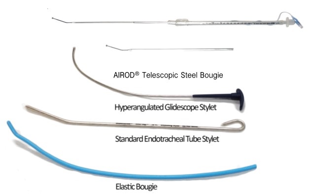
The choice of the correct endotracheal introducer can make the difference between first-pass success and failure (Figure 5).

Figure 5. Types of airway introducers.

The standard endotracheal tube stylet is used most often during direct laryngoscopy. This stylet may be bent when used with a curved Macintosh blade or without a bend when used with a straight Miller blade. The former is the most common method. An elastic bougie has an advantage over the standard stylet as it can be placed through the vocal cords and into the trachea, allowing better access especially with anterior airways during direct laryngoscopy with a Macintosh or Miller blade.

The BEAM (Bougie Use in Emergency Airway Management) trial is attracting renewed interest in intubation with a bougie rather than a stylet (2). In the BEAM trial, first-attempt success using an elastic bougie was compared to a stylet during laryngoscopy in an emergency department.

First-attempt success was achieved in 98% of patients compared to 87% in all patients. In patients with at least one difficult airway characteristic, first-attempt success using an elastic bougie was 96% compared to 82% using a stylet.

In the First-Attempt Endotracheal Intubation Success Rate Using a Telescoping Steel Bougie (3), intubation first-attempt success rate was 97% in the ICU. Subgroup analysis of first-attempt intubation success using the AIROD® to intubate in patients with a difficult airway was 96%.

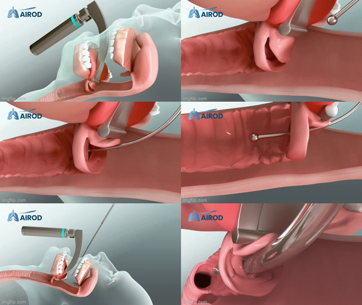
The average time to intubate was 15 seconds. During multiple intubations, the AIROD® was used to lift the epiglottis and move excess oropharyngeal tissue, improving the view of the glottis without causing any trauma to the airway (Figure 6).

Figure 6. Video of AIROD® lifting the epiglottis.

The hyperangulated Glidescope® stylet can be used with the Glidescope®, curved Macintosh blade, and C-MAC® blade. The AIROD® can be used with any direct or video laryngoscopy in any configuration: curved, hyperangulated, or straight.

The elastic bougie cannot make the acute turn required with hyperangluated laryngoscopes and should be avoided with this device unless the hyperangulated Glidescope® stylet is placed first and becomes caught up on the superior angle of the vocal cords. If this occurs, leave the Glidescope® in position and gently remove the hyperangulated Glidescope® stylet. While maintaining the acute angle, introduce an elastic bougie into the ETT and advance the tip into the trachea. Then slide the ETT down the bougie and into the trachea. 

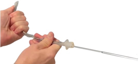
An alternative is to use the AIROD® steel bougie from the beginning, along with the Glidescope®. Load an ETT from the bulbous tip of the AIROD®, then shape to accommodate airway anatomy (Figures 7 and 8).

Figure 7. AIROD® shaped to accommodate airway anatomy.

Figure 8. ETT advancing down the AIROD®.

Use the proximal tip to lift the epiglottis and expose the vocal cords. Then advance the AIROD® two cm into the trachea followed by the ETT.

Case Presentations

Case 1

54-year-old man with severe coronary artery disease on aspirin and Plavix® with a history of a seizures associated with alcohol withdrawal became unresponsive and a code blue was called. He was found to be apneic with oxygen saturation in the 50s. He was stimulated by the hospitalist and became responsive. He was transferred to the ICU, where he became completely unresponsive again and stopped breathing. He was immediately ventilated with a bag-valve mask, and oxygenation improved to 100%. He then bolted up out of bed and became very combative. Propofol was given and he was laid supine and ventilated with a bag-valve mask. Inspection of his oropharynx revealed a very large tongue, and some missing and multiple sharp teeth with mouth opening of only 2 fingerbreadths. There was blood and emesis in his oropharynx that was suctioned. A Miller 4 blade was inserted into the oropharynx but only a grade 4 view could be obtained. The AIROD® was inserted into the oropharynx in the fully extended and locked position and the proximal tip was used to gently lift the epiglottis, exposing the vocal cords, and improving the view to a grade 2. The AIROD® was advanced 2 cm past the vocal cords and an assistant advanced an 8.0 endotracheal tube down the AIROD® until it was grasped, and the endotracheal tube was advanced successfully past the vocal cords while the assistant held the distal end of the AIROD®. The AIROD® was removed intact without any oropharyngeal or vocal cord trauma.

Case 2

A 63-year-old  5’5 110 kg woman with COPD, morbid obesity, obstructive sleep apnea, atrial fibrillation, diabetes mellitus, and anxiety suffered a cardiac arrest and was successfully resuscitated with placement of a drug eluting stent into the right coronary artery. One week later she required intubation for acute respiratory failure. She was extubated the following day and developed stridor, which resolved with pain medication and racemic epinephrine. Two days later, she developed acute respiratory failure again, with stridor that resolved after receiving 4 mg IV Versed. A diagnosis of paroxysmal vocal cord dysfunction was made. The next day she developed similar symptoms that responded to additional Versed® and Precedex®. The next morning, she became anxious after the Precedex® was stopped and once again developed acute stridor with respiratory failure, responding to Zyprexa® and Versed® momentarily. She was comfortable throughout the day until her stridor resumed, and despite BiPAP she was unable to adequately ventilate. She became obtunded, prompting intubation.

In addition to stridor, her Mallampati was 4, she had a sharp, prominent full set of teeth, an airway opening 1.5 cm, a large tongue with excessive oropharyngeal tissue, false cords, and vocal cord swelling. The AIROD® was preloaded with a 7.0 ETT that had attached to it a 10 mL syringe onto the distal end and tucked it under the patient’s right shoulder with the tip lying flat and pointing laterally, protected with a sterile OR towel. The AIROD® lay at a 45o to the neck. She was given 20 mg of etomidate and immediately ventilated with a bag-valve mask. A Miller 4 blade was gently inserted into the mouth, revealing a grade 4 view with purulent mucus in her oropharynx. The AIROD® was grasped and used to manipulate the false cords, revealing the true vocal cords while cricoid pressure was applied. A grade 2 view was obtained. The cords were adducted with a posterior glottal chink. The AIROD® was gently passed 2 cm through the tiny opening at the bottom of the vocal cords and used to dilate the area with the smooth bulbous tip. The ETT was then advanced into the trachea while the respiratory therapist held the distal end of the AIROD®. The AIROD® was removed intact without any evidence of oropharyngeal trauma. Successful first-attempt intubation occurred without complications. Bronchoscopy confirmed no tracheobronchial tree trauma.

Case 3

A 71-year-old  5’10’’ tall 101 kg man with non-insulin dependent diabetes mellitus, hypertension, and obesity was intubated 18 days prior for severe ARDS secondary to SARS-CoV-2. He subsequently lost his airway, and the attending physician was unable to intubate using the Glidescope®; so an emergency tracheostomy was performed with placement of a 5.0 Shiley. The evening of the 24th day of ventilation, he was unable to be ventilated effectively with his PCO2 rising to 73 mmHg with a pH of 7.13. He was on a propofol drip and 10 mg vecuronium was given while he was being ventilated through the 5.0 tracheostomy. He was actively bleeding from his nasopharynx. A Miller 4 blade was gently inserted into his mouth revealing a bloody and swollen oropharynx. A pre-loaded AIROD® was used to gently displace tissue, revealing a grade 1 view. The AIROD® was inserted 1 cm past the vocal cords and the ETT was then advanced slowly into the trachea with no assistant holding the AIROD®. The AIROD® was pulled back as the endotracheal tube was advanced down the trachea, abutting the tracheostomy tube. The ETT balloon was inflated and the AIROD® was removed intact without any evidence of acute oropharyngeal trauma. The single-handed first-attempt intubation was performed in 19 seconds. This was followed by the exchange of the 5.0 tracheostomy for an 8.0 tracheostomy. Bronchoscopy confirmed no acute oropharyngeal or tracheal trauma with the tracheostomy in the correct position in the trachea.

Case 4

A 68-year-old 5’10 126 kg smoker with a past medical history significant for COPD, on home oxygen with multiple intubations in the past was admitted. He had a past medical history of  pulmonary embolism on Eliquis®, deep venous thrombosis with an inferior vena cava filter, obstructive sleep apnea, and obesity. He was diagnosed with COVID-19 pneumonia and treated with BiPAP at 100% FiO2 for six days in the ICU. He developed ARDS and altered mental status, prompting intubation. Obese, large neck with limited neck mobility, micrognathia, large very dry tongue, sharp teeth with some missing, and a mouth opening 2 cm. He received propofol 200 mg IV and succinylcholine 200 mg IV. A Miller 4 blade gently inserted into oropharynx revealed an anterior glottis with false cords. The AIROD® was used to probe the false cords and advanced gently 5 cm, feeling the tracheal rings to ensure placement in the trachea. An 8.0 ETT was slowly advanced into the trachea using the single-handed first-attempt technique. An endotracheal balloon was inflated and the AIROD® removed intact without any evidence of acute oropharyngeal or tracheal trauma.

Case 5

28-year-old 5’9 man 97 kg with a past medical history significant for alcoholism was admitted. He was currently drinking two liters of vodka daily, had a history of  alcoholic cardiomyopathy and esophageal varices, drank hand sanitizer “to remain drunk”, and developed acute shortness of breath, and felt that his “throat was closing”. He developed very severe stridor with respiratory distress and was transferred to the ICU. Audible stridor could be heard as he arrived. He was in severe respiratory failure, sitting up, and very anxious. He was drooling bloody secretions. He was placed on a 15 L/min 100% FiO2 non-rebreathing mask. He was obese, had a large large neck with limited mobility, mouth opening 2 cm, protruding large tongue, full set of teeth, micrognathia with severe stridor, and was barely moving any air. He was given 4 mg IV Versed®. A tracheostomy kit was at bedside with a surgeon present. He was given 100 mg IV propofol, then laid flat and quickly placed in the SNIFF position. Bag-valve-mask was performed. SpO2 100%. An additional 100 mg IV propofol was given. A Miller 4 blade barely lifted the tongue when fresh blood was encountered. The blade was advanced gently, and bloody secretions suctioned. A crowded anterior hamburger oropharynx, bleeding with mucosal sloughing and false cords was encountered. The AIROD® pre-loaded with a 6.5 ETT was gently advanced underneath the epiglottis and advanced 3 cm, followed by advancement of the 6.5 ETT. Bag-valve ventilation occurred with poor CO2 detector color change. The ETT was left in place while bag-valve-mask ventilation was performed. SpO2 100%. The AIROD® was pre-loaded with a 7.0 ETT. A second-attempt revealed an air bubble anterior to the ETT. The 6.5 ETT was removed as the AIROD® was advanced towards the air bubble. The AIROD® was used to probe the hamburger glottis and to peel back the false cords revealing a small view of the right vocal cords, followed by advancement of the AIROD® 3 cm. A 7.0 ETT was slowly advanced into the trachea and balloon inflated with no assistant holding the AIROD®. No evidence of acute oropharyngeal trauma. Bronchoscopy revealed no tracheobronchial trauma and confirmed acute adenoviral necrotizing pharyngitis.

Conclusion

Anticipation of a difficult airway should always be considered, and having the necessary tools available can improve first-attempt endotracheal intubation success. Optimizing head positioning can be performed quickly and will help with glottic exposure. Knowing how to use multiple laryngoscopes as well as introducers can make the difference between life and death.

Conflicts of Interest

Evan D. Schmitz, MD is the inventor of the AIROD® and CEO of AIROD Medical, LLC.

Acknowledgments

The author thanks H. Carole Schmitz and Bille J. Maciunas for their editorial comments.

References

  1. Sasano N, Morita M, Sugiura T, Sasano H, Tsuda T, Katsuya H. Time progression from the patient's operating room entrance to incision: factors affecting anesthetic setup and surgical preparation times. J Anesth. 2009;23(2):230-4. [CrossRef] [PubMed]
  2. Driver B, Prekkar M, Klein L, et al. Effect of use of a bougie vs endotracheal tube and stylet on first-attempt intubation success among patients with difficult airways undergoing emergency intubation a randomized clinical trial. JAMA. 2018;319(21):2179-2189. [CrossRef] [PubMed]
  3. Schmitz ED. Decreasing COVID-19 patient risk and improving operator safety with the AIROD during endotracheal intubation. J of Emergency Services. EMSAirway. 11/2020.
  4. Adnet F, Borron SW, Dumas JL, Lapostolle F, Cupa M, Lapandry C. Study of the "sniffing position" by magnetic resonance imaging. Anesthesiology. 2001 Jan;94(1):83-6.[CrossRef] [PubMed]
  5. Adnet F, Baillard C, Borron SW, et al. Randomized study comparing the "sniffing position" with simple head extension for laryngoscopic view in elective surgery patients. Anesthesiology. 2001 Oct;95(4):836-41. [CrossRef] [PubMed]
  6. Schmitz ED, Park K. First-Attempt Endotracheal Intubation Success Rate Using A Telescoping Steel Bougie. Southwest J Pulm Crit Care. 2021;22(1):36-40. doi: [CrossRef]
  7. Schmitz ED, Park K. Emergency intubation of a critically ill patient with a difficult airway and avoidance of cricothyrotomy using the AIROD®. J of Emergency Medical Services. 2021;22(1):36-40. 
  8. Schmitz ED. Single-use telescopic bougie: case series. Southwest J Pulm Crit Care. 2020;20(2):64-8. [CrossRef]
  9. Schmitz ED. AIROD® Case Series: A New Bougie for Endotracheal Intubation. J of Emergency and Trauma Care. 2020;5(2):22.
  10. Schmitz ED. The Importance of Head Positioning During Endotracheal Intubation. EMSAirway. Jul 27, 2021. Available at: https://emsairway.com/2021/07/27/the-importance-of-head-positioning-during-endotracheal-intubation/#gref (accessed 4/18/23).
  11. Lascarrou JB, Boisrame-Helms J, et al. Video Laryngoscopy vs Direct Laryngoscopy on Successful First-Pass Orotracheal Intubation Among ICU Patients: A Randomized Clinical Trial. JAMA. 2017;317(5):483-493. [CrossRef] [PubMed]
  12. Higgs A, McGrath BA, Goddard C, Rangasami J, Suntharalingam G, Gale R, Cook TM; Difficult Airway Society; Intensive Care Society; Faculty of Intensive Care Medicine; Royal College of Anaesthetists. Guidelines for the management of tracheal intubation in critically ill adults. Br J Anaesth. 2018 Feb;120(2):323-352. [CrossRef] [PubMed]
  13. Brown CA 3rd, Bair AE, Pallin DJ, Walls RM; NEAR III Investigators. Techniques, success, and adverse events of emergency department adult intubations. Ann Emerg Med. 2015 Apr;65(4):363-370.e1. [CrossRef] [PubMed]
Cite as: Schmitz ED. Essentials of Airway Management: The Best Tools and Positioning for First-Attempt Intubation Success. Southwest J Pulm Crit Care Sleep. 2023;26(4):61-69. doi: https://doi.org/10.13175/swjpccs015-23 PDF
Wednesday
Mar012023

March 2023 Critical Care Case of the Month: A Bad Egg

Richard A. Robbins, MD

Phoenix Pulmonary and Critical Research and Education Foundation

Gilbert, AZ

History of Present Illness

You are asked to see a 35-year-old man who was admitted to the ICU from the ER the previous night with an exacerbation of his chronic obstructive pulmonary disease (COPD). He has a long history of COPD and came to the ER for COVID-19 testing because he was at a party where a friend was later found to COVID-19. He denies any change in his chronic respiratory symptoms but his spirometry was significantly worse than his baseline in the ER and despite his protests he was admitted. He was treated with empiric antibiotics (amoxicillin and clavulanic acid), corticosteroids (methylprednisolone 125 mg every 6 hours), bronchodilators (albuterol/ipratropium every 4 hours) and oxygen. He says his breathing has not improved and he wants to go home. He has had gradually increasing shortness of breath for the past 8-10 years. He has minimal cough but denied any fevers, systemic symptoms, or wheezing.  

PMH, FH, and SH

He had a history of multiple pneumothoraces which eventually led to bilateral pleurodesis. He has had not pneumothoraces since. He had a benign bone tumor removed about 25 years ago and a history of manic-depression. There is no FH of any similar type of problems. He does smoke about 3/4 pack of cigarettes per day and has more than occasional marijuana use.

Physical Exam

Physical examination was unremarkable expect for a well-healed scar on the left thigh.

Spirometry

Previous spirometry performed as an outpatient showed his FVC 2.54 L (53% of predicted) with an FEV1 1.25 L (31% of predicted). These improved to 2.99 L and 1.52 L after a bronchodilator. His spirometry last night in the ER was FVC 1.63 L (29 % predicted) and FEV1 0.80 L (18 % predicted).

Radiography

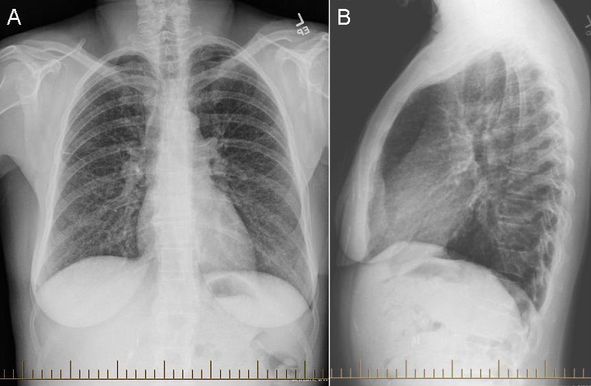
A chest radiograph was performed (Figure 1).

Figure 1. PA (panel A) and lateral (panel B) chest x-ray.

What should be done at this time? (Click on the correct answer to be directed to the second of five pages)

  1. Continue his antibiotics, corticosteroids and bronchodilators
  2. Order an alpha-1 antitrypsin level
  3. Transfer to the floor
  4. 1 and 3
  5. All of the above
Cite as: Robbins RA. March 2023 Critical Care Case of the Month: A Bad Egg. Southwest J Pulm Crit Care Sleep. 2023;26(3):28-30. doi: https://doi.org/10.13175/swjpccs009-23 PDF 
Tuesday
Jan242023

The Effect of Low Dose Dexamethasone on the Reduction of Hypoxaemia and Fat Embolism Syndrome After Long Bone Fractures

Dr. Akash K

Dr. Madhuchandra R

Department Of Orthopaedics, Karnataka Institute Of Medical Sciences, Hubli, India

Abstract

Background: A dangerous and sometimes fatal consequence of post-traumatic long bone fractures is fat embolism syndrome (FES). The reported incidence of FES ranges from 2% to 22%. FES can also lead to critical illness with fatality rates between 10 to 36%. This study's objective was to determine whether prophylaxis of the fat emboli syndrome could be achieved with lower doses of dexamethasone than had previously been recommended. Thus, prevention of respiratory insufficiency and disruption of homeostasis are essential.

Methods: A total of 583 adult cases of long bone shaft fracture patients between January 2020 to December 2021 were randomly divided into a trial group (n= 252) and a control group (n=331) by simple randomization. The trial group received dexamethasone 8mg/day for 3 days and the control group was given placebo. FES was diagnosed using Gurd’s diagnostic criteria and the FES morbidity and death rates in each group were examined.

Results: Five patients (0.151%) in the control group and 1 patient (0.39%) in the trial group developed FES but the difference was not significant (p>0.05). SpO2 values were significantly elevated in the dexamethasone-treated group compared to the control group 24 hours after admission (p<0.05) and the elevation persisted on the third post admission day (p<0.05).

Conclusion: Dexamethasone in low doses reduces post-traumatic hypoxia in patients with long bone fracture. However, our study was underpowered to show a reduction in FES.

Introduction

Fat emboli occur in all long bone fractures with the most severe resulting in fat embolism syndrome (FES). The reported incidence of FES ranges from 2% to 22% with fatality rates of 10-36% (1-3) with FES resulting in the adult respiratory distress syndrome a 50–90% mortality rate (1-3). Unfortunately, this is particularly common in young people in their second and third decades of life who sustain polytrauma and/or femur fractures in high-velocity traffic accidents (2,3). The majority of trauma patients may experience a subclinical form of FES, which manifests as  hypoxaemia alone (3-6).

FES resulting in systemic symptoms is a rare clinical outcome. Following a traumatic incident, fat droplets are released into the bloodstream resulting in fat embolization. This results in immediate tissue damage as well as a systemic inflammatory response that produces symptoms in the lungs, skin, nervous system, and retina (7,8). Most instances of FES occur after trauma but rare cases of FES have been reported to occur after bone marrow transplantation, osteomyelitis, pancreatitis, alcoholic fatty liver, and even liposuction (9,10). Although the classic triad of pulmonary distress, mental status changes, and petechial rash is usually not seen, hypoxia 24 to 48 hours after pelvic or long-bone fractures is common (11-13).

FES has no pathognomonic characteristics and laboratory and radiographic findings are nonspecific (14,15). Early detection of FES may allow supportive pulmonary treatment and other life-saving interventions to stop the pathophysiologic cascade and stop clinical deterioration. The majority of curative methods created expressly for FES have failed (16,17). There have been several attempts to avoid FES since it is such a serious issue in trauma patients (4). With varying degrees of success, heparin, dextran, albumin, hypertonic glucose, aspirin, and early fracture stabilization, have all been attempted (4). Steroids have also been studied as a preventative as well as a therapeutic agent in fat embolism in various studies.

When fat droplets act as emboli and are trapped in the pulmonary microvasculature and other microvascular beds, such as the brain, they may cause clinical symptoms to appear 24-72 hours after trauma (and particularly after fractures). Embolization starts out very slowly and reaches its peak in 48 hours or more. A long-acting corticosteroid having a half-life of 36 to 72 hours is dexamethasone. This study's objective was to determine whether prophylaxis of the fat emboli syndrome could be achieved with lower doses of dexamethasone than had previously been recommended (17).

Patients and Methods

From January 2020 to December 2021, 583 adult patients between the ages of 18 and 60 with long bone fractures without a history of chronic heart, lung, liver, or renal failure were recruited from patients at KIMS Hospital Hubli. There were 211 cases observed in women and 372 cases in men. The injuries resulted from motor accidents (426), falls (127), and crush injuries (30). Fracture sites included 128 femur fractures, 285 tibia and fibula fractures, 79 humerus fractures, and 91 pelvic injuries. The patients were randomized into two groups, one receiving dexamethasone and the other receiving a placebo (Table 1).

Table 1. Demographic data
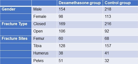
Click here to display Table 1 in a separate, enlarged window.

The following patient information was recorded: gender, age, weight, time from injury to admission, primary fracture location, type of fracture, FES morbidity, and number of fatalities. All patients received traditional medical care, early hypovolemic shock correction, fracture stabilization, and symptomatic therapy (2). The trial group received dexamethasone 8mg/day for 3 days and the control group was given placebo. All patients were monitored (heart rate, BP, SpO2 ,respiratory rate, urine output, and arterial blood gases) every 6 hours for 3 days. We considered hypoxaemia with any pO2 <70mm Hg and classified all patients in 3 categories; severe (pO2<60mm Hg), mild hypoxaemia (pO2 >60- <70 mm Hg) and normal (pO2>70mm Hg). All patients signed an informed consent form. The study was approved by the Ethics Committee of our institute hospital.

Treatment and diagnosis for FES

Patients were identified using “Gurd’s Diagnostic criteria score(Table 2), and those whose score was 2 major or 1 major and 4 minor were diagnosis as FES.

Table 2. Gurd’s Diagnostic Criteria Score*
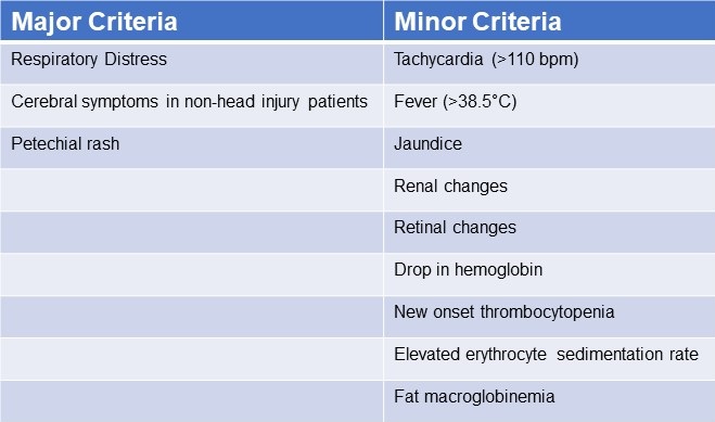
*Two major criteria or 1 major criterion and 4 minor criteria suggest a diagnosis of FES. Click here to view Table 2 in a separate and enlarged window.

Data analysis

Utilizing statistical tools, the analysis was conducted (SPSS 20.0). P< 0.05 was regarded as statistically significant when comparing the patients' age, main fracture location, fracture type, and incidence of FES using the chi-squared test and single-factor analysis of variance, respectively.

Results

FES occurred in the dexamethasone group and control group, with 1 and 5 cases, respectively (Table 3). Statistical analysis revealed that there was no statistically significant difference between the groups for sex, age, weight, injury to admission time, main fracture site, fracture type, or medication time.

Table 3. Incidence of FES

Click here to view Table 3 in a separate, enlarged window.

Twenty-four hours after admission, steroid treated patients displayed a statistically significant higher PaO2 value compared to the control group (p<0.05) and this difference persisted through the 3rd post admission day (p<0.05, table 4).

Table 4. Partial pressures of oxygen (in mm Hg) in patients treated with IV dexamethasone and controls.

Click here to view Table 4 in a separate and enlarged window.

Discussion

Much higher dosages of dexamethasone have been used to treat some pathological conditions in order to reduce inflammation, inhibit the immune system, impact the hemopoietic system, and alter metabolism (18-28). The mechanical-chemical hypothesis of fat embolism hypothesizes that neutral triglycerides are hydrolyzed into glycerol and free fatty acids by lipoprotein lipase from the lungs. The free fatty acids lead to inflammation and endothelial damage. Corticosteroids likely act on FES by reducing this inflammation. Due to a lack of clear diagnostic markers, treating FES may prove challenging. There have been few publications on the use of adrenal steroids to prevent high-risk FES patients, although the results have been ambiguous at low doses (31). Observational clinical research revealed that short-range and high doses may be helpful in reducing plasma free fatty acid concentrations, maintaining PaO2 levels, and reducing the occurrence of long bone fractures in individuals with FES. Dexamethasone may be a more effective drug treatment for FES (32). The dose of dexamethasone used in our study was relatively small and short, and complications related to hormones such as stress ulcer, aseptic necrosis of the femoral head, and bleeding tendency did not occur. It should be noted that drug prevention must be based on early, accurate fracture fixation, early corrective hypovolemic shock, and other standard procedures (33). This is true even if drug usage in this population clearly has a preventative impact. Ashbaugh and Petty (34) suggested corticosteroid therapy for treating FES in 1966 and gave laboratory data proving its therapeutic impact in the experimental animal given an intravenously administered FFA injection. Rokkanen et al. (35) found that 5 mg/kg of dexamethasone administered at 1 and 48 h after burn injury failed to enhance nuclear translocation of the GR, and to suppress the overproduction of proinflammatory cytokines such as TNF-α and IL-1β, neither did it increase the release of anti-inflammatory cytokine IL-10. In experiments with animals, Kreis et al. (36) showed that corticosteroids increased oxygenation and lowered the pathological alterations seen in lung biopsies. Alho et al. (37) conducted research on the use of intravenous methyl prednisolone sodium succinate in the prevention of fat embolism syndrome. A total of 60 individuals with at least two fractures were included in his study (pelvic, femoral or tibial fractures).methyl prednisolone reduces signs of  hypoxaemia, bilateral "snow storm" infiltrations of the lungs, petechial rash, mental disturbances, pyrexia, anemia and thrombocytopenia. Varying degrees of the syndrome were observed in two patients given methylprednisolone and in 15 patients in the control group. Babalis et al. (39) results support the prophylactic administration of methylprednisolone in small dosage to prevent post traumatic hypoxaemia and probably FES in patients with isolated lower limb long bone fractures, especially when early fracture stabilization is not possible. Therefore, every study has demonstrated the effectiveness of steroids as a preventative treatment for the fat embolism syndrome.

Although our results showed a trend towards reduction in FES after long bone fractures, the results were not statistically significant. This is likely because our study turned out to be underpowered. We had anticipated an incidence of FES between 2-20% reported in the literature rather than the 1.1% found in our study.

Conclusion

The study's objective was to determine whether prophylaxis of the fat emboli syndrome could be achieved with lower doses of dexamethasone than had previously been recommended. Among the several prophylactic drugs that have been researched so far for the fat embolism syndrome, dexamethasone have shown to be relatively beneficial. The frequency of  hypoxaemia and fat emboli syndrome decreased with intravenous dexamethasone at 8 mg per day for three days. Dexamethasone is a long-acting symptoms that emerge 24-72 hours after trauma (and particularly after fractures). Fat embolization begins slowly and reaches its maximum around 48 hours.

The limitation of our study is that it lacked sufficient power to demonstrate a reduction in FES. Furthermore, no method has been developed to pinpoint precisely who could benefit from steroid prophylaxis. We based our study assuming an incidence of FES of about 5%. However, we found an incidence of only about 1.5%. The lower incidence is probably due to our use of Gurd’s criteria which is more restrictive than the criteria used in other studies. Based on our observed incidence of FES of 1.5% with a reduction to 0.4% we estimate that over 2500 patients would be needed to show a statistically significant reduction in FES.

Our study shows that  hypoxaemia is reduced by a relatively low dose of dexamethasone administered for a relatively short length of time. It may prevent FES but our study was underpowered to show a difference.

Declaration

Human subjects: Consent was obtained or waived by all participants in this study. Karnataka Institute Of Medical Sciences ethics committee. issued approval 327/2020-21. The study was approved by the institutional ethics committee. Animal subjects: All authors have confirmed that this study did not involve animal subjects or tissues. Conflicts of interest: In compliance with the ICMJE uniform disclosure form, all

authors declare the following: Payment/services info: All authors have declared that no financial support was received from any organization for the submitted work. Financial relationships: All authors have declared that they have no financial relationships at present or within the previous three years with any organizations that might have an interest in the submitted work

References

  1. Sproule BJ. Brady JL. Gilbert J. Studies on the Syndrome of Fat Embolization. Can Med Assoc J. 1964 May 30;90(22):1243-7. [PubMed]
  2. Wertzberger JL, Peltier LF. Fat embolism: the importance of arterial hypoxia. Surgery. 1968 Apr;63(4):626-9. [PubMed]
  3. Stürm JA, Lewis FR Jr, Trentz O, Oestern HJ, Hempelman G, Tscherne H. Cardiopulmonary parameters and prognosis after severe multiple trauma. J Trauma. 1979 May;19(5):305-18. [CrossRef] [PubMed]
  4. Hutchins PM, Macnicol MF. Pulmonary insufficiency after long bone fractures. Absence of circulating fat or significant immunodepression. J Bone Joint Surg Br. 1985 Nov;67(5):835-9. [CrossRef] [PubMed]
  5. Levy D. The fat embolism syndrome. A review. Clin Orthop Relat Res. 1990 Dec;(261):281-6. [PubMed]
  6. Gossling HR, Pellegrini VD Jr. Fat embolism syndrome: a review of the pathophysiology and physiological basis of treatment. Clin Orthop Relat Res. 1982 May;(165):68-82. [PubMed]
  7. Kwiatt ME, Seamon MJ. Fat embolism syndrome. Int J Crit Illn Inj Sci. 2013 Jan;3(1):64-8. [CrossRef] [PubMed]
  8. Parisi DM, Koval K, Egol K. Fat embolism syndrome. Am J Orthop (Belle Mead NJ). 2002 Sep;31(9):507-12. [PubMed]
  9. Scuderi CS. The present status of fat embolism. Bibliographic review. Int Surg Digest 1934; 18: 195-215.
  10. Gurd AR. Fat embolism: an aid to diagnosis. J Bone Joint Surg Br. 1970 Nov;52(4):732-7. [PubMed]
  11. Nixon JR, Brock-Utne JG. Free fatty acid and arterial oxygen changes following major injury: a correlation between  hypoxaemia and increased free fatty acid levels. J Trauma. 1978 Jan;18(1):23-6. [CrossRef] [PubMed]
  12. Parker FB Jr, Wax SD, Kusajima K, Webb WR. Hemodynamic and pathological findings in experimental fat embolism. Arch Surg. 1974 Jan;108(1):70-4. [CrossRef] [PubMed]
  13. Nijsten MW, Hamer JP, ten Duis HJ, Posma JL. Fat embolism and patent foramen ovale. Lancet. 1989 Jun 3;1(8649):1271. [CrossRef] [PubMed]
  14. Vedrinne JM, Guillaume C, Gagnieu MC, Gratadour P, Fleuret C, Motin J. Bronchoalveolar lavage in trauma patients for diagnosis of fat embolism syndrome. Chest. 1992 Nov;102(5):1323-7. [CrossRef] [PubMed]
  15. White T, Petrisor BA, Bhandari M. Prevention of fat embolism syndrome. Injury. 2006 Oct;37 Suppl 4:S59-67. [CrossRef] [PubMed]
  16. Laterre PF, Wittebole X, Dhainaut JF. Anticoagulant therapy in acute lung injury. Crit Care Med. 2003 Apr;31(4 Suppl):S329-36. [CrossRef] [PubMed]
  17. Bederman SS, Bhandari M, McKee MD, Schemitsch EH. Do corticosteroids reduce the risk of fat embolism syndrome in patients with long-bone fractures? A meta-analysis. Can J Surg. 2009 Oct;52(5):386-93. [PubMed]
  18. McEvoy GK, Snow EK, Kester L, eds. AHFS 2002 Drug Information. Bethesda, MD: American Society of HealthSystem Pharmacists; 2002.
  19. Chamberlain D. Emergency medical treatment of anaphylactic reactions. Project Team of the Resuscitation Council (UK). J Accid Emerg Med. 1999 Jul;16(4):243-7. [CrossRef] [PubMed]
  20. Niermeyer S, Kattwinkel J, Van Reempts P, et al. International Guidelines for Neonatal Resuscitation: An excerpt from the Guidelines 2000 for Cardiopulmonary Resuscitation and Emergency Cardiovascular Care: International Consensus on Science. Contributors and Reviewers for the Neonatal Resuscitation Guidelines. Pediatrics. 2000 Sep;106(3):E29. [CrossRef] [PubMed]
  21. Brun-Buisson C, Brochard L. Corticosteroid therapy in acute respiratory distress syndrome: better late than never? JAMA. 1998 Jul 8;280(2):182-3. [CrossRef] [PubMed]
  22. Hudson LD. New therapies for ARDS. Chest. 1995 Aug;108(2 Suppl):79S-91S. [CrossRef] [PubMed]
  23. Meduri GU, Headley AS, Golden E, Carson SJ, Umberger RA, Kelso T, Tolley EA. Effect of prolonged methylprednisolone therapy in unresolving acute respiratory distress syndrome: a randomized controlled trial. JAMA. 1998 Jul 8;280(2):159-65. [CrossRef] [PubMed]
  24. Johnson MJ, Lucas GL. Fat embolism syndrome. Orthopedics. 1996 Jan;19(1):41-8; discussion 48-9. [CrossRef] [PubMed]
  25. Kallenbach J, Lewis M, Zaltzman M, Feldman C, Orford A, Zwi S. 'Low-dose' corticosteroid prophylaxis against fat embolism. J Trauma. 1987 Oct;27(10):1173-6. [PubMed]
  26. Niewoehner DE, Erbland ML, Deupree RH, Collins D, Gross NJ, Light RW, Anderson P, Morgan NA. Effect of systemic glucocorticoids on exacerbations of chronic obstructive pulmonary disease. Department of Veterans Affairs Cooperative Study Group. N Engl J Med. 1999 Jun 24;340(25):1941-7. [CrossRef] [PubMed]
  27. Richards RR. Fat embolism syndrome. Can J Surg. 1997 Oct;40(5):334-9. [PubMed]
  28. Kubota T, Ebina T, Tonosaki M, Ishihara H, Matsuki A. Rapid improvement of respiratory symptoms associated with fat embolism by high-dose methylpredonisolone: a case report. J Anesth. 2003;17(3):186-9. [CrossRef] [PubMed]
  29. Han YY, Sun WZ. An evidence-based review on the use of corticosteroids in peri-operative and critical care. Acta Anaesthesiol Sin. 2002 Jun;40(2):71-9. [PubMed]
  30. Habashi NM, Andrews PL, Scalea TM. Therapeutic aspects of fat embolism syndrome. Injury. 2006 Oct;37 Suppl 4:S68-73. [CrossRef] [PubMed]
  31. Babalis GA, Yiannakopoulos CK, Karliaftis K, Antonogiannakis E. Prevention of posttraumatic hypoxaemia in isolated lower limb long bone fractures with a minimal prophylactic dose of corticosteroids. Injury. 2004 Mar;35(3):309-17. [CrossRef] [PubMed]
  32. Yamamoto T, Irisa T, Sugioka Y, Sueishi K. Effects of pulse methylprednisolone on bone and marrow tissues: corticosteroid-induced osteonecrosis in rabbits. Arthritis Rheum. 1997 Nov;40(11):2055-64. [CrossRef] [PubMed]
  33. Talbot M, Schemitsch EH. Fat embolism syndrome: history, definition, epidemiology. Injury. 2006 Oct;37 Suppl 4:S3-7. [CrossRef] [PubMed]
  34. Ashbaugh DG, Petty TL. The use of corticosteroids in the treatment of respiratory failure associated with massive fat embolism. Surg Gynecol Obstet. 1966 Sep;123(3):493-500. [PubMed]
  35. Rokkanen P, Alho A, Avikainen V, Karaharju E, Kataja J, Lahdensuu M, Lepistö P, Tervo T. The efficacy of corticosteroids in severe trauma. Surg Gynecol Obstet. 1974 Jan;138(1):69-73. [PubMed]
  36. Kreis WR, Lindenauer SM, Dent TL. Corticosteroids in experimental fat embolization. J Surg Res. 1973 Mar;14(3):238-46. [CrossRef] [PubMed]
  37. Alho A, Saikku K, Eerola P, Koskinen M, Hämäläinen M. Corticosteroids in patients with a high risk of fat embolism syndrome. Surg Gynecol Obstet. 1978 Sep;147(3):358-62. [PubMed]
  38. Stoltenberg JJ, Gustilo RB. The use of methylprednisolone and hypertonic glucose in the prophylaxis of fat embolism syndrome. Clin Orthop Relat Res. 1979 Sep;(143):211-21. [PubMed]
  39. Babalis GA, Yiannakopoulos CK, Karliaftis K, Antonogiannakis E. Prevention of posttraumatic hypoxaemia in isolated lower limb long bone fractures with a minimal prophylactic dose of corticosteroids. Injury. 2004 Mar;35(3):309-17. [CrossRef] [PubMed]
Cite as: K A, R M. The Effect of Low Dose Dexamethasone on the Reduction of Hypoxaemia and Fat Embolism Syndrome After Long Bone Fractures. Southwest J Pulm Crit Care Sleep. 2023;26(1):11-17. doi: https://doi.org/10.13175/swjpccs059-22 PDF
Saturday
Nov192022

Unintended Consequence of Jesse’s Law in Arizona Critical Care Medicine

Adrienne Lee Jones-Adamczyk, RN, MSN, ACNP-BC, ACHPN, HEC-C

Patricia Ann Mayer, MD, MS, HEC-C

Banner Gateway Medical Center

Gilbert, AZ USA


Abstract

Jesse’s Law, passed in Arizona as a reaction to a surrogate acting against the interests of a specific patient, now prevents intensivists and surrogates who are acting appropriately from discontinuing unwanted interventions in dying hospice patients. The law prohibits statutory surrogates from authorizing discontinuance of artificial nutrition and hydration unless they can present “clear and convincing evidence” to a court that the patient would agree. This law is causing undue harm to hospice patients at end of life by delaying withdrawal of unwanted medical interventions, interfering with accepted and established surrogate decision-making precepts, and negating informed consent because surrogates are unaware that artificial nutrition and hydration cannot be easily discontinued after initiation. The authors offer a case example followed by an ethical analysis of this presumably unintended consequence of the law.

Abbreviations

  • ANH:     artificial nutrition and hydration
  • ICU:       intensive care unit
  • LST:       life sustaining treatment
  • PVS:      persistent vegetative state
  • SDM:     surrogate decision maker
  • TBI:       traumatic brain injury

Unintended Consequence of Jesse’s Law in Arizona Critical Care Medicine

We present a composite but common case demonstrating an unfortunate result of Jesse's Law affecting intensivists and their patients who are at end of life. We follow with a short history and discussion of the ethical implications of the law.

Case Report: An 88 y/o widowed woman was admitted to an intensive care unit (ICU) in Arizona in respiratory failure after driving herself to the local emergency department. By the time her family was reached, she was intubated, on dialysis, and had a feeding tube placed for artificial nutrition and hydration (ANH). 

Over the next several days, she worsened and developed multi-organ failure. In conversations with the family, the intensivist elucidated that the patient lived alone and generally declined to complain or seek medical help. The family relayed she was a third-generation Arizonan who had grown up on the family ranch, where she still lived. She'd often told her family: "When my time comes, it comes; don't keep me alive on machines and tied to tubes. If I'm on my way out, just take me home and let me go." Like many patients, she lacked written advanced directives, but her extended family as her surrogates agreed "her time had come." They requested removal of all tubes and machines and discharge to the ranch with hospice services and family in attendance. As the orders were being written, the nurse asked the intensivist: "What about Jesse's law? We can't just take the feeding tube out and stop the feeding." The nurse was correct. This makes little sense for our patient.

How and why did Arizona get here?

In May of 2007, 36-year-old Jesse Ramirez and his wife were involved in a rollover car crash reportedly caused by a heated argument between the two. Jesse suffered a severe traumatic brain injury (TBI) and was in a coma. Ten days later, his wife, as his statutory surrogate, chose to move him to hospice and discontinue his ANH. Jesse's siblings filed suit, contending that his wife did not have his best interests at heart given their severe marital discord. The Arizona court ruled in favor of Jesse's siblings, and his tube feedings were continued. He moved from hospice to rehab and later regained some function including the ability to recognize and interact with his family (1). Jesse's law, prohibiting surrogates from discontinuing ANH, was passed in 2008 as a reaction to this unfortunate case.

Jesse's Law states: "There is a rebuttable presumption that a patient who does not have a valid living will, power of attorney or other health care directive has directed the patient's health care providers to provide the patient with food and fluid to the degree that is sufficient to sustain life, including, if necessary, through a medically invasive procedure… and … that provision is in the patient's best interests.”(2) The law, therefore, allows only a legally appointed medical power of attorney or a court-appointed guardian but not a statutory surrogate to discontinue ANH for non-medical reasons. The law listed no exceptions, which meant the critical care team could not discontinue our patient’s feeding tube unless her surrogate decision makers (SDMs) obtained permission from a court.

How does Jesse’s law align with the national evolution of patient rights at end-of-life? It doesn’t.

Those rights, including withdrawing and withholding life-sustaining treatment (LST), date to 1976 with the Karen Ann Quinlan case (3). Karen Ann suffered an anoxic brain injury following a respiratory arrest and was subsequently determined to be in a persistent vegetative state (PVS). When months passed without improvement, her family requested the discontinuation of her ventilator based on their belief that Karen Ann would not want her life prolonged in her current condition. The hospital and her treating physicians initially denied this request fearing accusations of murder. The case eventually reached the New Jersey Supreme Court, which allowed removal of the ventilator and set two groundbreaking precedents. First, the Court determined that families are appropriate SDMs for incapacitated patients. Second, the Court determined patients and SDMs do have the right to refuse LST (4).

The second major case, that of Nancy Cruzan, began in 1990. (3) This young woman's parents as her SDMs also requested withdrawal of LST, but in this case, the LST was her feeding tube. Nancy was also in a PVS after a car accident but did not need a ventilator; she had been kept alive through ANH alone. Nancy’s case was the first withdrawal of ANH to be heard by the US Supreme Court. Although the ruling was multifaceted, it did allow withdrawal of the feeding tube, and Nancy died eight years after her accident once her ANH was discontinued (5).

These landmark cases clearly established SDMs as appropriate medical decision-makers for incapacitated patients and empowered them to withhold or withdraw medical treatments, including ANH (3). Along with this power, SDMs have the obligation to make decisions according to accepted criteria, namely 1) the wishes of the patient 2) if patient wishes are unknown, then SDMs are to use substituted judgment, that is, to make the decision they believe the patient would make if she were able to speak for herself or 3) in the absence of the first two, SDMs are to act in the patient’s best interest (2,6).

Jesse’s law in Arizona creates an exception to these precedents. Although Arizona allows withholding or withdrawing other LST by SDMs (including ICU treatments), it does not allow for the withdrawal of ANH, even when the SDM has clear knowledge of the patient's wishes (2). Jesse's law specifically presumes that a patient receiving ANH who lacks advance directives wants – in all cases - to prolong life and continue ANH indefinitely without regard to prognosis, quality of life, or verbalized preferences as told to SDMs (2). This includes the patient described in our case, who clearly would not have wanted continued ANH as she was dying.

Jesse's law, with its lack of exceptions, therefore, causes undue harm at the end of life for dying Arizona patients because it makes assumptions about patient wishes and conflicts with patient autonomy. The law focuses on ANH when the real problem in Jesse’s case was an SDM who was clearly not acting in his best interests. Although young patients with brain injuries like Jesse may recover over time, our terminally ill patient could not; yet the law prohibited the ICU team from removing (withdrawing) her feeding tube.

Indeed, withholding and withdrawing LST have long been considered ethically equivalent. (3,7,8,9). McGee (7) reports stopping (withdrawing) ANH is akin to an omission (withholding). The accepted ethical premise is that omissions do not cause death; actions do. Therefore stopping ANH is no more a cause of death than not starting ANH would be. Similarly, Beauchamp and Childress (8), the founders of principles in modern medical ethics, assert no morally relevant difference between ANH and other types of LST types. They add the "right to refuse treatment should not be contingent on the type of treatment" offered. The American Academy of Neurology agrees and openly opposes legislation that presumes to know a patient's wishes regarding ANH and/or limits the ability of patients to declare their preferences, including through discussions with SDMs (9). Current ethical consensus supports an appropriately acting SDM (not the case with Jesse’s wife) to authorize withholding or withdrawing ANH as well as to make any other medical decision a surrogate would make.

Unfortunately, Jesse's law interferes with both autonomy and the informed consent process in Arizona for dying patients. Respect for autonomy allows patients (or their SDMs) to accept or reject recommended medical treatments that affect their bodies. "Every person being of adult years and sound mind has the right to determine what shall be done with his own body"(10). Respect for autonomy includes a requirement of informed consent. In Arizona, SDMs who consent to ANH do not then have the authority to withdraw consent unless they go to court to present "clear and convincing evidence" that the patient would refuse ANH (2). Few SDMs are aware of this when ANH is started, fewer still have the time or energy for a court appearance when faced with a dying loved one. And since informed consent requires the SDM to have "adequate and truthful information about the risk versus benefits and understand the treatment goals", we posit consent is often not obtained regarding ANH for patients such as ours (6).

An informed consent conversation for ANH includes at least three key points (6,11):

1) ANH is a medical treatment and not a basic intervention…for all patients;

2) ANH provides uncertain benefits for many diagnoses and has considerable risks and discomfort;

3) ANH is not a comfort measure since symptoms associated with not eating or drinking can be palliated and generally resolve within a short period of time.

We add that, in Arizona, the ANH informed consent conversation with surrogates ought to specify that permission for ANH cannot be withdrawn (without court intervention) once given. 

ANH is rarely indicated for patients with a terminal illness at end-of-life. It carries significant risks, including bleeding, infection, aspiration, and the use of physical or chemical restraints to prevent a patient from dislodging the required tubes. There is no evidence that ANH at the end of life leads to improved survival or quality of life; it is rarely beneficial and often harmful (7,11). And yet, Jesse's law makes no easy provisions for such patients.

Our patient wanted to die unencumbered by medical interventions including her feeding tube, but the ICU team could not accommodate that request under current Arizona law. So, what choices remain for our patient, her surrogates and the ICU team? The team can leave the feeding tube in place, or the surrogates can try and convince a court to allow its removal, spending time in court instead of with their loved one.

We assert that Jesse's law, with its lack of exceptions for patients such as ours, creates undue distress and barriers for intensivists and surrogates attempting to honor patient wishes and end ANH appropriately in dying patients. Jesse’s law should have addressed unreasonable surrogates instead of preventing all surrogates from taking an action that is often in the best interest of a loved one.

Conflict of Interest Statement: The authors have no conflict of interest and nothing to disclose. Both authors are employees of Banner Health.  

References

  1. Grado G. (2011, October 8). Crash survivor's case spurs new state law. East Valley Tribune. Retrieved September 15, 2022, from https://www.eastvalleytribune.com/local/crash-survivor-s-case-spurs-new-state-law/article_486772f1-4bd0-59e3-b24f-073bbce7b543.html AZ HB2823.
  2. AZ HB2823. Fifty-fifth Legislature 1st Regular. (2021, May 24). LegiScan. Retrieved November 07, 2022, from https://legiscan.com/AZ/bill/HB2823/2021
  3. Fine RL. From Quinlan to Schiavo: medical, ethical, and legal issues in severe brain injury. Proc (Bayl Univ Med Cent). 2005 Oct;18(4):303-10. [CrossRef] [PubMed]
  4. In re Quinlan, 70 N.J. 10, 355 A.2d 647 (NJ 1976)
  5. Cruzan v. Director, Missouri Department of Health, 110 S. Ct. 2841 (1990), 19 Fla. St. U. L. Rev. 209 (1991).
  6. Shah P, Thornton I, Turrin D, Hipskind  J. (2022, June 11). Informed Consent. Stat Pearls. Retrieved November 8, 2022, from https://www.ncbi.nlm.nih.gov/books/NBK430827/
  7. McGee A. Does withdrawing life-sustaining treatment cause death or allow the patient to die? Med Law Rev. 2014 Winter;22(1):26-47. [CrossRef] [PubMed]
  8. Beauchamp TL, Childress JF, Principles of Biomedical Ethics. 8th ed. New York, NY: Oxford University Press; 2019: 165-166.
  9. Bacon D, Williams MA, Gordon J. Position statement on laws and regulations concerning life-sustaining treatment, including artificial nutrition and hydration, for patients lacking decision-making capacity. Neurology. 2007 Apr 3;68(14):1097-100. [CrossRef] [PubMed]
  10. Schloendorff v. New York Hospital, 211 N.Y. 125, 105 N.E. 92 (N.Y. 1914).
  11. Casarett D, Kapo J, Caplan A. Appropriate use of artificial nutrition and hydration--fundamental principles and recommendations. N Engl J Med. 2005 Dec 15;353(24):2607-12. [CrossRef] [PubMed]
Cite as: Jones-Adamczyk AL, Mayer PA. Unintended Consequence of Jesse’s Law in Arizona Critical Care Medicine. Southwest J Pulm Crit Care Sleep. 2022;25(5):83-87. doi: https://doi.org/10.13175/swjpccs051-22 PDF