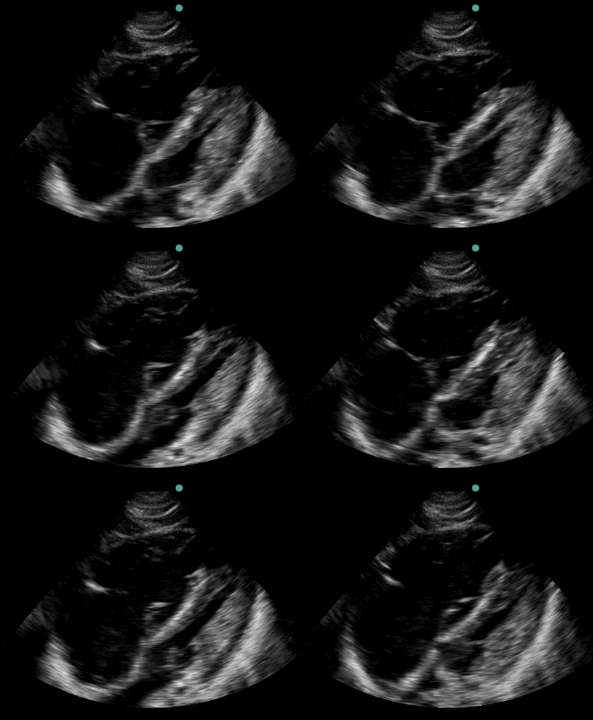
A 32 year old man was admitted a week earlier with sickle cell pain crisis. He had developed increasing dyspnea, oxygen desaturation and bilateral pulmonary infiltrates. He had a pulseless electric activity code blue and an ultrasound of the heart was obtained (Figure 1).

Figure 1. Subxiphoid view ultrasound of the heart.
What does the ultrasound show?
- Aortic dissection
- Aortic stenosis
- Enlarged left ventricle
- Enlarged right ventricle
- Pericardial effusion
Reference as: Raschke RA. Ultrasound for critical care physicians: sickle cell crisis. Southwest J Pulm Crit Care. 2013:7(2):110-1. doi: http://dx.doi.org/10.13175/swjpcc113-13 PDF