Search Journal-type in search term and press enter
Southwest Pulmonary and Critical Care Fellowships
In Memoriam

Critical Care Journal Club

(Click on title to be directed to posting, most recent listed first)

November 2017 Phoenix Critical Care Journal Club
Tucson Critical Care Journal Club: Albumin Use in the Critical Care Unit
May 2016 Phoenix Critical Care Journal Club: Oxygen Therapy
November 2015 Tucson Critical Care Journal Club: Atrial Fibrillation in
   Sepsis
September 2015 Phoenix Critical Care Journal Club: Goal-Directed
Therapy and DNR
June 2015 Phoenix Critical Care Journal Club: Interventions in ARDS
February 2015 Phoenix Critical Care Journal Club: Intracranial Pressure
Monitoring for Fulminant Liver Failure
August 2014 Tucson Critical Care Journal Club: Bacteremia in Cardiac 
   Arrest
July 2014 Phoenix Critical Care Journal Club: Predicting the Future
April 2014 Tucson Critical Care Journal Club: Early Goal-Directed
   Therapy
April 2014 Phoenix Critical Care Journal Club: Early Goal-Directed
   Therapy
March Critical Care Journal Club: Sequelae of Critical Care
February 2014 Phoenix Critical Care Journal Club: Subgroup Analysis
January 2014 Critical Care Journal Club
November 2013 Critical Care Journal Club
September 2013 Banner Good Samaritan / Phoenix VA Critical Care
   Journal Club
September 2013 Tucson Critical Care Journal Club: Early
   Tracheostomy
August 2013 Critical Care Journal Club: Less is More
July 2013 Critical Care Journal Club
April 2013 Critical Care Journal Club
March 2013 Critical Care Journal Club
February 2013 Critical Care Journal Club
January 2013 Critical Care Journal Club
December 2012 Critical Care Journal Club
November 2012 Critical Care Journal Club
October 2012 Critical Care Journal Club
September 2012 Critical Care Journal Club
August 2012 Critical Care Journal Club
July 2012 Critical Care Journal Club
June 2012 Critical Care Journal Club
May 2012 Critical Care Journal Club
April 2012 Critical Care Journal Club
February 2012 Critical Care Journal Club
January 2012 Critical Care Journal Club
December 2011 Critical Care Journal Club 
November 2011 Critical Care Journal Club
September 2011 Critical Care Journal Club 
August 2011 Critical Care Journal Club 
July 2011 Critical Care Journal Club
June 2011 Critical Care Journal Club 
April 2011 Critical Care Journal Club
March 2011 Critical Care Journal Club
February 2011 Critical Care Journal Club
November 2010 Critical Care Journal Club
October 2010 Critical Care Journal Club


Both the Phoenix Good Samaritan/VA and the University of Arizona fellows previously had a periodic critical care journal club in which current or classic articles were reviewed and discussed. A brief summary was written of each discussion describing the article and the strengths and weaknesses of each article.

-------------------------------------------------------------------------------------

Friday
Apr252014

April 2014 Phoenix Critical Care Journal Club: Early Goal-Directed Therapy 

The ProCESS investigators. A randomized controlled trial of protocol-based care for early septic shock.  New Engl J Med. 2014 epub ahead of print. Available at: http://www.nejm.org/doi/full/10.1056/NEJMoa1401602 (accessed 4/24/14).

Editor's note: The April 2014 Tucson Critical Care Journal Club also reviewed this article and interpreted the data somewhat differently.

We were fortunate to be joined in our discussion by Dr. Frank LoVecchio, one of the primary investigators of the ProCESS trial, and doctors Robbins, Bajo, Mand and Thomas, as well as our pulmonary critical care fellows.

The ProCESS trial was important for two reasons: first, it showed that early goal-directed therapy (EGDT) does not benefit patient mortality; second, it provides another example of how the evidence-based practice of critical care medicine has often been misguided by invalid evidence. In this aspect, EGDT joins the ranks of tight glucose control, drotrecogin alpha (Xigris®), Swan Ganz catheter-guided resuscitation, corticosteroids, and other interventions in our field that were once part of evidence-based practice, but ultimately found to lack benefit or even be harmful to our patients. That recurrent theme in our literature is the main point of this Journal Club.

The first example of an algorithm for goal-directed therapy (GDT) that we found in the literature was written by Shoemaker in 1975 (1). Shoemaker’s proposed algorithm is surprisingly modern-appearing and similar to River’s EGDT algorithm. Resuscitation with IVF, blood, vasopressors, and vasodilators (dobutamine was just being developed for commercial use in the 1970s) was aimed at achieving specific goals for blood pressure, central venous pressure, pulmonary artery wedge pressure and hematocrit. Even in the 1960’s, it was known that CVP was not a reliable goal for fluid management though, and Shoemaker continued to refine his algorithm, steering away from a CVP-based approach.  

Shoemaker published a very influential paper in 1988 (2). This small RCT showed a dramatic mortality benefit of goal-directed therapy aimed at achieving a supranormal cardiac output and oxygen delivery monitored by a Swan Ganz catheter. This approach was based on the theory that resuscitation should not just normalize, but optimize oxygen delivery to tissues, thereby increasing tissue oxygen consumption and preventing organ failure. “Delivery-dependent oxygen consumption” was a theory that made very good sense – and took a decade to disprove.

Delivery-dependent oxygen consumption-based GDT algorithms were subsequently extensively researched and implemented over the next decade. As residents and junior faculty in the late 80s and early 90s, we got lots of experience putting in Swans and calculating oxygen delivery to guide resuscitation efforts. But to our great disappointment sequential studies failed to confirm any benefit of our efforts. One of the studies we reviewed was an RCT with 762 patients that demonstrated resuscitation focused on a goal of achieving a mixed venous oxygen saturation > 70% in critically ill patients provided no mortality benefit (3). By 2003, an RCT with nearly 2000 patients that showed no benefit of Swan-based GDT in high-risk surgical patients (4) was the final nail in the coffin of what was beginning to be called “the Cult of the Swan-Ganz” – referring to those of us who had trouble giving up our faith in the value of pulmonary artery wedge pressures and oxygen delivery calculations. It was eventually shown that the entire theoretical basis for Swan-based GDT was invalid – that oxygen consumption by tissues was independent of oxygen delivery, except when oxygen delivery got very, very low.  Several studies suggested that Swan-Ganz catheters might actually increase patient mortality.

It may have been hard for the fellows, who didn’t share this experience, to understand the skepticism that seasoned clinicians had regarding River’s “EGDT” trial published in 2001 (5). There was little new about River’s version of GDT except the location of care in the emergency room. The goals of EGDT were the same as those that were for the most part tested and disproven decades earlier. There was nothing new therapeutically – IVF, blood, pressors and inotropes had all been part of GDT algorithms since the 1980s. Yet River’s trial showed a dramatic reduction of mortality from 46.5% to 30.5% (p=0.009, NNT: 6.25) in patients presenting with severe sepsis or septic shock. River’s version of GDT became the backbone of evidence-based resuscitation bundles, recommended by the Surviving Sepsis Campaign, the Institute for Healthcare Improvement, and others. By 2008, many non-experimental studies had been published that all seemed to support River’s original study results, and greatly expand it’s clinical generalizability outside the ER. In fact, the pooled risk reduction when a meta-analysis was performed by Rivers himself was actually even better than in his original study – with a NNT of 5 for every life saved (6). Rivers concluded that these cumulative findings were so incontrovertible, that future efforts should focus on “overcoming logistical, institutional, and professional barriers to implementation”. This opinion was widely shared and coordinated efforts were made to incentivize sepsis bundle compliance by physicians.

Before proceeding to the ProCESS trial, recall the setting in which it occurred, because we have been here many times before, and we will be here many times again. As a profession, we were convinced.  “Bundle compliance” was considered as failed if even a single aspect of EDGT was not achieved within the proper time frame. Those few who remained skeptical about EGDT had now become “professional barriers” to implementation of one of the most robust-appearing examples of evidence-based medicine in our literature. Yet the subsequent negative results of ProCESS ought not to have been a surprise when viewed in light of the history of evidence-based practice in Critical Care.

The randomized controlled trial of Protocol-based Care for Early Septic Shock (ProCESS) enrolled 1341 patients in 31 emergency rooms across the country. Patients were randomized to three groups: River’s-type EGDT, a simplified resuscitation protocol (in which central venous lines were only placed if needed to provide vasopressor infusion, and resuscitation goals were based on blood pressure and pulse), and a non-protocolized “usual care” group. Sixty-day mortality in the groups were 21%, 18% and 19% respectively – statistically non-significant. Not to lose sight of the forest for the trees, we will not further detail the study design of ProCESS, but it enrolled more than five times the number of patients as in River’s single-site trial, and there were no major flaws in the design or implementation.

ProCESS ought to teach us this: (again). Evidence-based interventions in critical care that originally appear to reduce mortality are almost always subsequently found to be of no benefit, or to actually harm the patient. This fundamental observation transcends how we normally look at evidence-based medicine. Normally, we take the most recent randomized controlled trial, and abide by the conclusions that we can draw from it. But this approach has repeatedly proven to be wrong. Of all major non-prophylactic treatment interventions in critical care medicine which have been shown to provide a mortality benefit, only ARDSnet low tidal volume ventilation has stood the test of time. Swans, steroids, Xigris, tight glucose control, EGDT, and possibly even post-arrest hypothermia have all fallen. We ought to learn some humility from this in regards to formulating and enforcing rigid protocols for patient care. 

More trial evidence is forthcoming. The ongoing Australasian Resuscitation in Sepsis Evaluation (ARISE – but I wonder why it’s not just “ARSE”), and Protocolised Management in Sepsis trial (ProMiSe) in the UK will likely shed more light (or more confusion) on the issue. But even if EGDT didn’t specifically work after all, the effect of River’s study continues to be important. On one hand, we have spent an enormous amount of effort and money trying to rigidly comply with a sepsis resuscitation bundle that is currently disproven to provide any benefit. On the other hand, it’s fascinating to note that control group mortality fell from 46% to 19% in the twelve years between River’s trial and ProCESS. This is an incredible success even if it’s only because we paid more attention to sepsis, and not because we complied with a specific resuscitation bundle. We have learned that early diagnosis of sepsis is essential. I hope we will also learn that we can make great strides in critical care without having to all be in lock-step with one particular protocol. There are likely many ways to provide good care for a critically-ill patient. It’s important to have a plan for how to do that, but it’s unlikely that any single plan is better than all others.

Robert A. Raschke, MD

Banner Good Samaritan Medical Center  

Phoenix, AZ

References 

  1. Shoemaker WC. Algorithm for resuscitation: a systematic plan for immediate care of the injured or postoperative patient. Crit Care Med. 1975;3:127-130. [CrossRef] [PubMed] 
  2. Shoemaker WC, Appel PL, Kram HB, Waxman K et al. Prospective trial of supranormal values of survivors as therapeutic goals in high-risk surgical patients. Chest. 1988;94:1176-86. [CrossRef] [PubMed] 
  3. Gattinoni L, Brazzi L, Pelosi P, Latini R. et al. A trial of goal-oriented hemodynamic therapy in critically ill patients. New Engl J Med. 1995;333:1025-33. [CrossRef] [PubMed] 
  4. Sandham JD, Hull RD, Brant RF, Knox L, et al. A randomized controlled trial of the use of pulmonary-artery catheters in high-risk surgical patients. New Engl J Med. 2003;348:5-14. [CrossRef] [PubMed] 
  5. Rivers E, Nguyen B, Havstad S, Ressler J, Muzzin A, Knoblich B, Peterson E, Tomlanovich M; Early Goal-Directed Therapy Collaborative Group. Early goal-directed therapy in the treatment of severe sepsis and septic shock. New Engl J Med. 2001;345:1368-77. [CrossRef] [PubMed] 
  6. Rivers E, Coba V, Whitmill M. Early goal-directed therapy in severe sepsis and septic shock: a contemporary review of the literature. Curr Opin Anesthesiol. 2008;21:128-140. [CrossRef] [PubMed] 
  7. The ProCESS investigators. A randomized controlled trial of protocol-based care for early septic shock.  New Engl J Med. 2014 epub ahead of print. Available at: http://www.nejm.org/doi/full/10.1056/NEJMoa1401602 (accessed 4/24/14).

Reference as: Raschke RA. April 2014 Phoenix critical care journal club: early goal-directed therapy. Southwest J Pulm Crit Care. 2014;8(4):239-42. doi: http://dx.doi.org/10.13175/swjpcc057-14 PDF

Friday
Apr112014

March Critical Care Journal Club: Sequelae of Critical Care

We focused on the topic of long-term sequelae of acute respiratory failure requiring mechanical ventilation.  

Our discussion panel included the fellows, many of our faculty including Drs. Robbins, Mathew, Singarajah, Thomas, Rinne, Garcia-Orr, and Nair, and invited guests from Palliative Care Medicine: Dr. Carleton, and Julie Lehn (from the VAMC and BGSMC respectively).

The long term clinical outcomes of two groups of patients were examined. The first group was comprised of survivors of acute respiratory distress syndrome (ARDS) requiring mechanical ventilation of any duration. Although the short-term mortality of ARDS has improved, previously unrecognized long-term sequelae have become a focus of research.  Studies have now shown that significant depression, cognitive deficits similar in magnitude to those of mild Alzheimer’s, and post-traumatic stress disorder (PTSD) each occur in approximately 25-30% of these patients (1-4). PTSD continues in about a quarter of patients even out to eight years after discharge (2). Functional disability is demonstrable up to 5 years after discharge, even in younger patients (4). Although pulmonary function typically normalize, all patients – even young patients with relatively mild ARDS – report subjective physical weakness out to five years post discharge (4). Twenty-seven percent of the patient’s family members will also suffer depression and PTSD in the aftermath of an ICU admission for ARDS.

The second group of patients are those who require mechanical ventilation for greater than 21 days, or who require tracheostomy. The term “Chronic Critical Illness” has been coined to describe this group of patients, who comprise 5-10% of ICU admissions (1). These patients are often “trached and PEGed” and transferred to skilled nursing facilities after ICU care is completed. It is estimated that there are approximately 100,000 such patients in the United States at any point in time. We all recognize the clinical manifestations that many of these patients share: persistent delirium, critical illness weakness, musculoskeletal catabolism, skin breakdown, dysphagia/anorexia/malnutrition, infection/colonization with healthcare-associated microorganisms, and prolonged renal failure, among others.

We often feel a sense of limited success when such a patient survives the ICU, but outcome studies suggest a very poor outlook for meaningful recovery (5,6). Less than half of these patients will ever be liberated from the ventilator, and almost never after more than 60 days of mechanical ventilation dependence. At 6 months, 75% will be dead or institutionalized. By a year, only about 10% will be living independently at home.  Chronically critically-ill patients with age >50, thrombocytopenia, and need for ongoing vasopressors or dialysis suffer 97% 1-year mortality. 84% of family caregivers of survivors end up having to quit their jobs or significantly cut back work hours in order to provide support - many of these family care givers suffer depression and physical health deterioration.  A $3.5 million dollar direct cost is incurred per independently functioning survivor of chronic critical illness at 1 year.  

We do not do a good job communicating these grim statistics to our patients and their families. Up to 90% of families of chronically critically ill patients report that they received no information about possible functional dependency or 1-year mortality at hospital discharge. Perhaps this is because it is hard for us to accept the futility of much of what we do. Besides our best efforts to minimize sedation, ambulate and extubate patients as rapidly as possible, and prevent iatrogenic infections, there is little of proven value to prevent or treat chronic critical illness.

Notwithstanding these depressing statistics there are things we can do to mitigate both the physical and psychological complications which result from the acute phase of the critical illness. The first, as noted above, is to simply take an interest in the chronic phase of critical illness by following through on the outcomes of our patients. It is well demonstrated that the actuarial age-and-sex adjusted survival for critical illness takes between two and five years to return to normal, depending on the disease process. It is shorter for surgical patients and longer for medical patients (who are frequently suffering the end result of chronic organ system failure, for which there is limited curability). The fragmentation of disposition in modern healthcare means that once these patients have left the intensive care unit their ongoing mortality and morbidity is invisible to us unless we take active steps to check in on their condition. It follows, incidentally, that it is not realistic to expect excellent survival statistics for long term acute care facilities when these institutions are taking from acute ICU’s the burden of a population predestined to have a high mortality and morbidity.

Intensive care follow up clinics, although almost non existent in the United States, largely for reasons of reimbursement, have become widespread in other parts of the world.  In the United Kingdom, for example, the National Health Service Initiative “Intensive care without walls” was launched in 2000. During this relatively early phase of development, research has been quite plentiful and has largely consisted of characterizing the problems described above. Earlier diagnosis of psychological sequelae such as post-traumatic stress disorder, cognitive impairment, depression, sleep derangement and also the physical sequelae such as deconditioning and chronic pain have allowed earlier outpatient treatment-which has, unsurprisingly, been shown to improve outcomes. However, with increasing rapidity management of the “back end” of critical illness-the chronic disease state- is moving closer and closer to the “front end” as we realize quite how tightly they are connected.

The first report of mobilization in mechanically ventilated patients was published only in 2007 but since then a steady series has begun to appear in the literature (7). In 2009 Schweikert et al (8) demonstrated that early physical and occupational therapy during ventilation resulted in a return to independent functional status at hospital discharge in 29 (59%) of patients compared with 19 (35%) patients in the control group (p=0.02; odds ratio 2.7). Moreover, ICU delirium days were reduced by 50% (2.0 days vs. 4.0 days, P = .03) in spite of no differences in sedatives administered and more ventilator-free days (23.5 days, p=0.05) during the 28-day follow-up period than did controls. The early mobilization strategy led to a 1.7-fold increase in patients who were functionally independent when they left the hospital (59% vs. 35%, P = .02). Finally, more patients in the early mobilization group went directly home after hospitalization than in the control group (43% vs. 24%, P = .06). Of note, there was just one serious adverse event in 498 therapy sessions and this consisted of arterial oxygen desaturation to less than 80%.

In 2013 the BRAIN-ICU study mentioned above demonstrated that longer duration of delirium was associated with worse cognition and executive function scores at 3 and 12 months post discharge (4). It is increasingly clear that policies which result in minimization of total sedative dose not only reduce delirium but also promote early mobilization and better REM sleep. Normal fatigue from early mobilization results in the virtuous cycle of not only better natural REM sleep with less GABA agonist needs but thereby a reduced incidence of delirium. Less delirium is associated with better long term psychological outcomes and early mobilization is associated with better long term physical outcomes. These factors are intimately interconnected both between each other in the ICU and also with the long term outcomes once the patient goes home. It simply makes sense. While on the one hand we need to take ownership of the truly terrible long term course of critical illness, on the other we should embrace the possibility of doing something significant to improve it even though we can’t easily see it unless we start to look. At the present time, it is hiding in plain sight.

The ensuing discussion led us to several conclusions. We felt that in cases of acute respiratory failure requiring short-term mechanical ventilation, patients and their families should be advised of the high risk of persistent neurocognitive, psychological and functional disability and offered a venue for appropriate follow-up after discharge. Establishment of a Critical Care follow-up clinic is worth considering in this regard.

Further measures are required in cases of prolonged ventilator dependence. This starts with the recognition that tracheostomy is most often NOT a step towards recovery, but more likely a sign that the patient will never regain independent function again. In fact, the prognosis in patients with chronic critical illness is often worse than most forms of cancer in terms of mortality and quality of life.

In most patients who do not liberate from the ventilator within three weeks, a frank discussion of the grim long-term outcomes should ensue before tracheostomy is performed. It is likely that some patients and families might chose other alternatives, such as withdrawal of life support, if given an accurate picture of the poor chance they will survive or ever regain independence. Palliative care medicine consultation should be strongly considered in all such patients. The Society of Critical Care Medicine has a helpful brochure with information for patients and families in regard to Chronic Critical Illness available on their website. Good bedside care of patients with chronic critical illness and their families will likely reduce unnecessary suffering while reducing the cost of healthcare.

Robert A. Raschke, MD and Huw Owen-Reece, MD

Banner Good Samaritan Medical Center    

References

  1. Nelson JE, Cox CE, Hope AA, Carson SS. Chronic Critical Illness. Am J Respir Crit Care Medicine. 2010;182:446-454. [CrossRef] [PubMed]
  2. Davydow DS, Desai SV, Needham DM, Bienvenu J. Psychiatric morbidity in survivors of the acute respiratory distress syndrome: A systematic review. Psychosomatic Medicine. 2008;70:512-9. [CrossRef] [PubMed] 
  3. Herridge MS, Tansey CM, Matte A, Tomlinson G, et al. Functional disability 5 years after acute respiratory distress syndrome. NEJM. 2011;364:1293-1305. [CrossRef] [PubMed] 
  4. Pandharipande PP, Girard TD, Jackson JC, Morandi A, et al. Long-term cognitive impairment after critical illness. NEJM. 2013;369:1306-16. [CrossRef] [PubMed]
  5. Carson, SS, Garrett J, Hanson LC, Lanier J et al. A prognostic model for one-year mortality in patients requiring prolonged mechanical ventilation. Crit Care Medicine. 2008;36:2061-69. [CrossRef] [PubMed]
  6. Wright JC, Plenderleith L, Ridley SA. Long-term survival following intensive care: subgroup analysis and comparison with the general population. Anaesthesia. 2003;58(7):637-42. [CrossRef] [PubMed] 
  7. Bailey P, Thomsen GE, Spuhler VJ, et al. Early activity is feasible and safe in respiratory failure patients. Crit Care Med. 2007;351:139-145. [CrossRef] [PubMed]
  8. Schweickert WD, Pohlman MC, Pohlman AS, et al. Early physical and occupational therapy in mechanically ventilated, critically ill patients: a randomized controlled trial. Lancet. 2009; 373(9678):1874-82. [CrossRef] [PubMed] 

Reference as: Raschke RA, Owen-Reece H. March critical care journal club: sequelae of critical care. Southwest J Pulm Crit Care. 2014;8(4):229-32. doi: http://dx.doi.org/10.13175/swjpcc049-14 PDF

Friday
Feb212014

February 2014 Phoenix Critical Care Journal Club: Subgroup Analysis

Sun X, Ioannidis JP, Agoritsas T, Alba AC, Guyatt G. How to use a subgroup analysis: users' guide to the medical literature. JAMA. 2014;311(4):405-11. [CrossRef] [PubMed]

One of Dr. Raschke's pet peeves is unplanned subgroup analysis. In the September 2013 Banner Good Samaritan / Phoenix VA Critical Care Journal Club (1) he commented on an article by Hung et al. (2) that used a post hoc subgroup analysis. He felt strongly enough to write to the editor about why post hoc subgroup analysis should not be acceptable as a basis for scientific conclusions and his letter was published this month (3). Therefore, we have been on the lookout for a review article to discuss subgroup analysis and came across this timely publication in JAMA.  The authors cite a number of examples and provide 5 criteria to use when assessing the validity of subgroup analyses:

  1. Can chance explain the apparent subgroup effect;
  2. Is the effect consistent across studies;
  3. Was the subgroup hypothesis one of a small number of hypotheses developed a priori with the direction specified;
  4. Is there strong preexisting biological support;
  5. Is the evidence supporting the effect based on within- or between-study comparisons.

The first 4 criteria are applicable to individual studies or systematic reviews, the last only to systematic reviews of multiple studies. These criteria will help clinicians deciding whether to use subgroup analyses to guide their patient care but we are all in agreement that care must be used when using subgroup analysis to not overestimate an effect.

Geurts M, Macleod MR, Kollmar R, Kremer PH, van der Worp HB. Therapeutic hypothermia and the risk of infection: a systematic review and meta-analysis. Crit Care Med. 2014;42(2):231-42. [CrossRef] [PubMed]

After reviewing Sun's article in JAMA above, we were on the alert for an article doing subgroup analysis and this one was interesting. Briefly, the authors performed a systematic review and meta-analysis of randomized trials to examine the risk of infections in patients treated with hypothermia. Twenty-three studies were identified, which included 2,820 patients, of whom 1,398 (49.6%) were randomized to hypothermia. In patients treated with hypothermia, the prevalence of all infections was not increased (rate ratio, 1.21 [95% CI, 0.95–1.54]), but there was an increased risk of pneumonia and sepsis (risk ratios, 1.44 [95% CI, 1.10–1.90]; 1.80 [95% CI, 1.04–3.10], respectively).

After reviewing Sun's article above, we were all skeptical about concluding that hypothermia caused pneumonia or sepsis especially since the overall rate of infections did not increase. The effect was not consistent between studies, none of the studies other than this meta-analysis hypothesized the association a priori and the preexisting biological support appears to be weak. However, we agree with the authors' recommendation that future randomized trials of hypothermia should report infections, pneumonia and sepsis to see if the association holds up.

Schortgen F, Clabault K, Katsahian S, Devaquet J, Mercat A, Deye N, Dellamonica J, Bouadma L, Cook F, Beji O, Brun-Buisson C, Lemaire F, Brochard L. Fever control using external cooling in septic shock: a randomized controlled trial. Am J Respir Crit Care Med. 2012;185(10):1088-95. [CrossRef] [PubMed]

After coming across the article that hypothermia might be associated with pneumonia and sepsis, Dr. Raschke suggested reviewing this article which appeared about two years ago. In this multicenter randomized controlled trial, febrile patients with septic shock requiring vasopressors, mechanical ventilation, and sedation were allocated to external cooling (n=101) to achieve normothermia or no external cooling (n=99). Vasopressors were tapered to maintain the same blood pressure target in the two groups. The primary endpoint was the number of patients with a 50% decrease in baseline vasopressor dose after 48 hours. from 12 hours of treatment (54 vs. 20%; P<0.001) but not at 48 hours (72 vs. 61%). Shock reversal was significantly more common with cooling and day-14 mortality were significantly lower in the cooling group. Please note that although external cooling was used in both the above meta-analysis on infection and hypothermia and this study, the patient population in this study were septic patients and the goal was to correct fever but not induce hypothermia.

Overall this was a good study that would encourage the use of fever control in septic shock patients. However, aside from this specific group of patients it is unclear if achieving normothermia is beneficial.

van Diepen S, Reynolds HR, Stebbins A, Lopes RD, Džavík V, Ruzyllo W, Geppert A, Widimsky P, Ohman EM, Parrillo JE, Dauerman HL, Baran DA, Hochman JS, Alexander JH. Incidence and outcomes associated with early heart failure pharmacotherapy in patients with ongoing cardiogenic shock. Crit Care Med. 2014;42(2):281-8. [CrossRef] [PubMed]

It seems that β-blockers are being used for everything these days. Guidelines recommend β-blockers and renin-angiotensin aldosterone system blockers (ACE inhibitors including angiotensin receptor blockers and aldosterone antagonists) to improve long-term survival in hemodynamically stable myocardial infarction (MI) patients with a reduced left ventricular ejection fraction. This is a subgroup analysis of the TRIUMPH trial which was an international, double-blind, multicenter, randomized, placebo-controlled study that evaluated the effect of tilarginine acetate, a nitric oxide synthase inhibitor, in patients with MI complicated by cardiogenic shock that persisted despite successful infarct artery revascularization. The authors compared 30-day mortality in patients in 66 patients (27.5%) had either β blocker or ACE inhibitors administered within the first 24 hours after the diagnosis of cardiogenic shock. The observed 30-day mortality among the treated patients was higher (27.3% vs. 16.9%; p = 0.035). In further subgroup analysis, the mortality was higher in those treated with only a β blocker (33.3% vs. 16.9%, p = 0.017) but not among those only treated with ACE inhibitors (18.2% vs. 16.9%, p = 1.000).

Even though this was a subgroup analysis, this manuscript supports the concept that β blockers in MI complicated by cardiogenic shock are probably harmful despite guidelines that might be interpreted as encouraging their use.

Adhikari NKJ, Dellinger RP, Lundin S, Payen P, Vallet B, Gerlach H, Park KJ, Mehta S, Slutsky AS, Friedrich JO. Inhaled nitric oxide does not reduce mortality in patients with acute respiratory distress syndrome regardless of severity: systematic review and meta-analysis. Crit Care Med. 2014;42(2):404-12. [CrossRef] [PubMed]

I have always been skeptical that inhaled nitric oxide (NO) does much for the adult respiratory distress syndrome (ARDS) other than transiently improve oxygenation presumably by dilating pulmonary arteries to areas of good ventilation. This meta-analysis included 9 trials (n = 1,142 patients) of inhaled NO in ARDS patients. NO did not reduce mortality in patients with severe ARDS (p = 0.93; n = 329, six trials) or mild-moderate ARDS (p = 0.33; n = 740, seven trials). The authors tried to look at several subgroups but could find no group that NO appeared to improve survival.

Inhaled NO may have a role in transiently increasing oxygenation in the severely hypoxic patient but these patients appear to be relatively rare. A number of years of ago an editorial was written by Warren Zapol entitled "Nitric oxide in acute respiratory distress syndrome: it works but can we prove it?" (4). It appears the answer is NO. It may be time to bury the concept that inhaled nitric oxide has much of a therapeutic role in ARDS, other than the severely hypoxic patient.

Reade MC, Finfer S. Sedation and delirium in the intensive care unit. N Engl J Med. 2014;370(5):444-54. [CrossRef] [PubMed]

Sedation and delirium are "hot" topics in the critical care world. Overall, this is a good review. There are also guidelines on this topic from the Society of Critical Care medicine (5). Both this review article and the guidelines restate the obvious (delirium is associated with higher mortality, EEG should be monitored in patients with seizures, etc.) but it is surprising how little quality data is available. In the guidelines most of the recommendations fall into B and C categories.

Richard A. Robbins, MD

Editor

References

  1. Raschke RA. September 2013 banner good samaritan / phoenix va critical care journal club. Southwest J Pulm Crit Care. 2013;7(4):241-4. [CrossRef]
  2. Hung IF, To KK, Lee CK, Lee KL, Yan WW, Chan K, Chan WM, Ngai CW, Law KI, Chow FL, Liu R, Lai KY, Lau CC, Liu SH, Chan KH, Lin CK, Yuen KY. Hyperimmune IV immunoglobulin treatment: a multicenter double-blind randomized controlled trial for patients with severe 2009 influenza A(H1N1) infection. Chest. 2013;144(2):464-73. [CrossRef] [PubMed] 
  3. Raschke RA. Post hoc subgroup analysis. Chest. 2014;145:435. [CrossRef] [PubMed]
  4. Zapol WM. Nitric oxide in acute respiratory distress syndrome: it works but can we prove it? Crit Care Med. 1998;26(1):2-3. [CrossRef] [PubMed]
  5. Barr J, Fraser GL, Puntillo K, et al. Clinical practice guidelines for the management of pain, agitation, and delirium in adult patients in the intensive care unit. Crit Care Med 2013;41:263-306. [CrossRef] [PubMed]

Reference as: Robbins RA. February 2014 Phoenix critical care journal club: subgroup analysis. Southwest J Pulm Crit Care. 2014;8(2):128-21. doi: http://dx.doi.org/10.13175/swjpcc021-14 PDF

Friday
Feb142014

January 2014 Tucson Critical Care Journal Club: Esmolol in Septic Shock

Morelli A, Ertmer C, Westphal M, et al. Effect of heart rate control with esmolol on hemodynamic and clinical outcomes in patients with septic shock: a randomized clinical trial. JAMA. 2013;310(16):1683-91. [CrossRef] [PubMed]

During septic shock, catecholamines serve to elevate heart rate and maintain cardiac output as an adaptive response to low peripheral vascular resistance. However, elevated serum catecholamines and tachycardia are associated with poor outcomes in the intensive care unit (ICU) (1). This study hypothesized that β-blocker therapy (intravenous esmolol) titrated to control heart rate would enhance myocardial function and improve outcomes in patients with septic shock.

This single center, randomized, open label, parallel group study randomized 154 participants to either receive intravenous esmolol titrated to achieve a heart rate of 80-94 beats per minute (bpm) or standard of care. Randomization occurred after 24 hours of hemodynamic optimization, including appropriate volume resuscitation. Inclusion criteria included the presence of septic shock requiring norepinephrine, heart rate > 95 bpm and age > 18. Exclusion criteria included a heart rate < 95 at randomization, significant cardiac or valvular dysfunction and pregnancy. The primary outcome was the difference in heart rate as measured by the mean area under the curves (AUCs) during the first 96 hours of treatment. Secondary outcomes included norepinephrine requirements, cardiopulmonary and oxygen indices, safety endpoints, and 28-day mortality.

Baseline patient characteristics were similar between the two treatment groups. Of note, there was an unusually high rate of multidrug-resistant Klebsiella and Acinetobacter baumannii among both groups. SAPS II scores were calculated based on variables at the time of randomization and were similar between the groups.

Compared to the control group, the intervention group achieved a lower median AUC for HR (P < .001) with all intervention group participants meeting the heart rate goal of 80-94 bpm. The intervention group had a 28-day mortality of 49.4% vs. 80.5% in the control group (P <.001, hazard ratio, 0.392).  Intervention group median AUC for norepinephrine requirements were significant less (P < .003) without a corresponding decrease in mean arterial pressure. Median AUC was significantly higher in the intervention group compared to the control group for stroke volume index (P < .02), systemic vascular resistance index (P < .001), left ventricular stroke work index (P = .03), GFR (P < .001), and PaO2/FiO2 (P = .03). Lastly, the intervention group median AUC for fluid requirements were significantly less (P < .001).

Although this was a well-designed study, there are several limitations to consider. First, the study was nonblinded, raising concern for unconscious bias in delivery of care. Second, there was an unusually high rate of multidrug-resistant Klebsiella and Acinetobacter baumannii among the two study groups. This finding makes it difficult to extrapolate the data to our patient populations with significantly lower rates of multidrug-resistant infections. Third, the mortality rates of both groups were much higher than their calculated SAPS II scores would suggest. This may be accounted for by their incorrect methodology for calculating a SAPS II score. Fourth, as the author’s state, the large difference in mortality between groups does not exclude the possibility of a chance finding or contributions from unknown confounding factors. Lastly, this was a single center study. It is well documented in the statistical literature that single center studies show larger effect size (2,3) When single center studies are reproduced into multicenter studies, the effect size is often less and occasionally nonexistent. This does not however negate the importance of this study.               

This appropriately designed phase 2 trial of targeted heart rate control for volume resuscitated septic shock patients with persistent tachycardia demonstrated a large mortality benefit without obvious adverse effects. The use of beta blockade during septic shock represents an exciting and novel treatment modality that warrants additional study to confirm these results. In the interim, clinicians should carefully consider the merits of early adoption in appropriately selected patients. While no clear disadvantages were identified in this well-done, single-center trial, the literature would caution us not to overestimate the early findings of promising new treatments.

Christopher Strawter MD, Cristine Berry MD, Christian Bime MD and Joe Gerald PhD

University of Arizona

Tucson, AZ

References

  1. Singer M. Catecholamine treatment for shock—equally good or bad? Lancet. 2007;370(9588):636-7. [CrossRef] [PubMed]
  2. Dechartres A, Boutron I, Trinquart L, Charles P, Ravaud P. Single-center trials show larger treatment effects than multicenter trials: evidence from a meta-epidemiologic study. Ann Intern Med. 2011;155(1):39-51. [CrossRef] [PubMed]
  3. Ioannidis JPA. Why most discovered true associations are inflated. Epidemiology. 2008; 19(5):640-8. [CrossRef] [PubMed] 

Reference as: Strawter C, Berry C, Bime C, Gerald J. January 2014 Tucson critical care journal club: esmolol in septic shock. Southwest J Pulm Crit Care. 2014;8(2):108-9. doi: http://dx.doi.org/10.13175/swjpcc016-14 PDF

Thursday
Feb062014

January 2014 Critical Care Journal Club

Kamps MJ, Horn J, Oddo M, Fugate JE, Storm C, Cronberg T, Wijman CA, Wu O, Binnekade JM, Hoedemaekers CW. Prognostication of neurologic outcome in cardiac arrest patients after mild therapeutic hypothermia: a meta-analysis of the current literature. Intensive Care Med. 2013;39(10):1671-82. [CrossRef] [PubMed]

Cohort studies performed prior to the advent of therapeutic hypothermia had shown that severe deficits in motor response to painful stimuli, or deficits in certain cranial nerve reflexes, could be used to rule-out the possibility of meaningful neurological recovery in patients who did not regain consciousness within 48-72 hours after cardiac arrest.  However, subsequent reports have challenged the reliability of these findings in patients who received therapeutic hypothermia – some of whom recovered despite grim neurological findings.

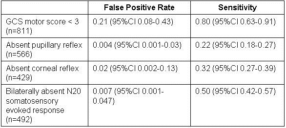
These authors performed a meta-analysis to determine whether neurological findings performed 72 hours after cardiac arrest (of all causes) could be used to prognosticate neurological outcome in patients who received mild therapeutic hypothermia.   They identified ten studies with a cumulative sample size of 1153 patients, and used this data to calculate the sensitivity and false positive rate for four neurological findings.  A poor neurological outcome was somewhat ill-defined, as included studies used different neurological scoring systems, and periods of follow-up varied, but generally would equate to an outcome no better than a vegetative state.  The following operating characteristics were calculated:

This study probably represents the best current data to support decisions regarding withdrawal of life support in patients who received therapeutic hypothermia post cardiac arrest.  A very low false positive rate is necessary to avoid recommending withdrawal of support from a patient who might recover.  Only absent pupillary response and absent N20 somatosensory evoked response meet this requirement.  Unfortunately, the sensitivity of these two tests are both poor, so we might expect that many will suffer prolonged life support to no good end if these tests are all we have to go on.  It would make the most sense clinically to look at the pupillary reflex first, then order the SSEP if the pupils react, but we have no information of the sensitivity of the tests used in series. 

The findings of this study will introduce more uncertainty into certain end-of-life discussions.  It has been posited that hypothermia might have confounded neurological prognostication simply by delaying clearance of sedation drugs.  If that is the case, future studies may show improved performance of clinical prognosticators by giving the patient more time to recover – perhaps waiting 72 hours after normothermia is achieved instead of 72 hours after the arrest.   Until further studies are available though, we will have to proceed as best we can in patients who do not awaken within 72 hours after cardiac arrest, taking into account the entire clinical status of each individual patient.  Younger patients with less comorbidity seem to have better recuperative capability.  Studies have shown that patients who suffer cardiac arrest secondary to sepsis or cancer have a very low chance of survival to discharge regardless of their neurological findings. 

Nielsen N, Wetterslev J, Cronberg T, et al. Targeted temperature management at 33°C versus 36°C after cardiac arrest. N Engl J Med. 2013;369(23):2197-206. [CrossRef] [PubMed]

Two previous randomized controlled trials have shown that patients who do not rapidly recover consciousness after cardiac arrest from ventricular fibrillation or ventricular tachycardia, have improved neurological outcomes if they receive 24 hours of therapeutic hypothermia in the range of 32-34C.  These authors set out to confirm this finding, and better define the optimal therapeutic temperature. 

They randomized 939 patients who remained unconscious after out-or-hospital cardiac arrest to temperature management with a goal temperature of 33C versus 36C.  Life support was discontinued during the first week in 247 patients based on neurological findings associated with poor outcomes.  These included brain death, status myoclonus, or a motor GCS score of two or less plus either status epilepticus or absent cortical N20 somatosensory evoked potential.  Mortality at the end of the study was 50% vs. 48% (p=0.51).  At 180-day follow-up, the percentage of patients who died or had poor neurological outcome was 54% vs. 52% (p=0.78).    

This study was well designed, and larger than both previous randomized controlled trials combined.  It was well powered to detect a 20% reduction in the hazard ratio for death.  180-day follow-up was accomplished for all survivors.  It seems to be the most convincing data available related to post-arrest hypothermia, but interpretation is debatable.  If the results are valid, it could mean that temperature control with a target of 36C is equally beneficial to temperature control with a target of 33C.  This would support continuation of our current practice of careful temperature management, with perhaps less danger of potential complications of hypothermia and less need for sedation to control shivering.  Alternatively, it could mean that therapeutic hypothermia doesn’t work after all.  The later interpretation might be consistent with a recent large RCT that showed that starting hypothermia therapy in the prehospital setting was not clinically beneficial in patients who suffered cardiac arrest, even though it significantly reduced the time it took to achieve a temperature of 34C. (JAMA 2014;311:45-52).  I think the first interpretation is more likely valid, and would recommend that we target 36C based on the findings of this study.  Therapeutic hypothermia is a labor-intensive and potentially dangerous therapy.  We have seen fatal hypothermia result from poor temperature control during a course of “therapeutic hypothermia”.  As with almost everything in our field, “less is more”. 

Slutsky AS, Ranieri VM. Ventilator-induced lung injury. N Engl J Med. 2013 Nov 28;369(22):2126-36. [CrossRef] [PubMed]

This paper did not address pulmonary complications of mechanical ventilation such as oxygen toxicity, or barotrauma, but it was a good overall review of ventilator associated lung injury (VALI).  The discussion of the interplay of ventilator pressures and volumes was particularly helpful. The main point here is that alveolar overdistention is likely the main mechanism of VALI.  Elevated transpulmonary pressure is closely related to lung overdistention, but it cannot be directly measured in any clinically practical way.  Transpulmonary pressure equals the alveolar pressure (plateau pressure) minus the pleural pressure.  We often use the alveolar pressure as a surrogate for transpulmonary pressure, but this can be misleading.  A morbidly obese patient on mechanical ventilation may require high alveolar pressure to inflate their heavy chest wall, without experiencing elevated transpulmonary pressures or overdistention.  Another patient in respiratory distress on a mechanical ventilator may have modest positive alveolar pressures, yet generate very high negative pleural pressures in their effort to pull breaths through the ventilator  – this could lead to elevated transpulmonary pressures and alveolar overdistention.   Therefore even though alveolar pressure (plateau pressure) was used in the ARDSnet low-tidal-volume RCT, it is not always an accurate measure of the risk for VALI.  Read the article for more details.

Cuenca AG, Gentile LF, Lopez MC, et al. Development of a genomic metric that can be rapidly used to predict clinical outcome in severely injured trauma patients. Crit Care Med. 2013;41(5):1175-85. [CrossRef] [PubMed]

This was a fantastic research question buried in a very hard-to-read paper with a weak study design.  The authors analyzed genomic data from a retrospective cohort of 167 patients who suffered severe trauma, and identified a panel of 63 leukocyte genes that distinguished between patients who had good and poor recoveries.  These were collected into a genomic panel that was then re-analyzed using a different laboratory method, and reduced to a single prognostic metric.  Although the authors call this stage of the study “validation”, it is not - since the same patients were simply retested for the same set of genes.   Still, the receiver operating characteristic curves shown in Figure 4 on page 1183 of the article are fascinating.  They show that APACHE II severity adjustment is almost without value in discriminating a complicated recovery in this set of patients, with an area-under-the-curve of 0.59 (a coin-toss would have an AUC of 0.50).  The genomic models had an AUC as high as 0.81.  The concept that we might be able to measure the response of our leukocytes to systemic injury, and that this response might be more predictive of outcomes than all the clinical elements of the APACHE II score is highly thought-provoking.  Hope to see a true validation study of this technology soon, using an independent sample of patients.     

Bernal W, Wendon J. Acute liver failure. N Engl J Med. 2013 Dec 26;369(26):2525-34. [CrossRef] [PubMed]

We have been interested in this topic for many years in relation to our liver transplant program, and our fellows and staff have previously developed and published a neuroprotective clinical protocol for the prevention and treatment of cerebral edema in patients with acute liver failure - the leading cause of death in ALF (1).   But we were disappointed in this review.  The authors offer only a brief treatment of the most controversial aspect of patient care – the use of intracranial pressure monitors.  The authors “monitor intracranial pressure only in patients with clinical signs or evidence of developing intracranial hypertension”, but do not discuss specifically what those are.  In fact, some clinical signs of intracranial hypertension, such as papilledema and Cushing reflex are notoriously insensitive for detecting the development of intracranial hypertension.  In our opinion, the most sensitive and practical clinical sign to detect intracranial hypertension is the development of grade III or IV encephalopathy.  The controversy regarding whether or how to best measure ICP is largely ignored in this article.  No mention is made of many important aspects of practical bedside management that are vital to supporting a patient through a period of intracranial hypertension.  For instance, no mention is made of the calculation and management of cerebral perfusion pressure (CPP) related to ICP monitoring.  [CPP=mean arterial pressure – ICP].   It is important to maintain adequate CPP (typically > 60 mmHg) during periods of intracranial hypertension in order to prevent ischemic brain injury.  We have also found head position to be strikingly important in patients with cerebral edema.  Patients with ICP monitors who are laid flat for central line placement often suffer immediate, potentially life-threatening increases in ICP.  This likely explains why ALF patients sometimes suffer brain injury during transport, or other situations in which their head may be lowered.  Prognostication is key to the decision to transplant the patient, or transport the patient to a transplant center – but the authors only address the topic in general terms, without actually providing any quantitative prognostic criteria or any specific operating characteristics of any of the competing prognostic scoring systems.  This review is inadequate for a critical care physician who actually provides bedside care of patients with acute liver failure.

Robert A. Raschke, MD MS

Associate Editor

References

  1. Raschke RA, Curry SC, Rempe S, Gerkin R, Little E, Manch R, Wong M, Ramos A, Leibowitz AI. Results of a protocol for the management of patients with fulminant liver failure. Crit Care Med. 2008;36(8):2244-8. [CrossRef] [PubMed]

Reference as: Raschke RA. January 2014 critical care journal club. Southwest J Pulm Crit Care. 2014;8(2):101-4. doi: http://dx.doi.org/10.13175/swjpcc012-14 PDF